[English] 日本語
Yorodumi
- PDB-2gza: Crystal structure of the VirB11 ATPase from the Brucella Suis typ... -

+
Open data


ID or keywords:

Loading...

-
Basic information

Entry
Database: PDB / ID: 2gza
Title
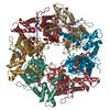
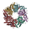
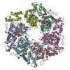
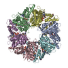
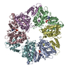
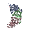
Crystal structure of the VirB11 ATPase from the Brucella Suis type IV secretion system in complex with sulphate
ComponentsType IV secretion system protein VirB11
KeywordsHYDROLASE / Secretion / ATPase
Function / homology
Function and homology information


secretion by the type IV secretion system / type IV secretion system complex / : / ATP binding / cytoplasm
Similarity search - Function
Type IV secretion system protein VirB11 / Beta-Lactamase - #90 / Type II/IV secretion system protein / Type II/IV secretion system protein / Sigma-54 interaction domain, ATP-binding site 1 / Beta-Lactamase / P-loop containing nucleotide triphosphate hydrolases / P-loop containing nucleoside triphosphate hydrolase / Rossmann fold / 2-Layer Sandwich ...Type IV secretion system protein VirB11 / Beta-Lactamase - #90 / Type II/IV secretion system protein / Type II/IV secretion system protein / Sigma-54 interaction domain, ATP-binding site 1 / Beta-Lactamase / P-loop containing nucleotide triphosphate hydrolases / P-loop containing nucleoside triphosphate hydrolase / Rossmann fold / 2-Layer Sandwich / 3-Layer(aba) Sandwich / Alpha Beta
Similarity search - Domain/homology
Type IV secretion system protein VirB11
Similarity search - Component
Biological speciesBrucella suis (bacteria)
MethodX-RAY DIFFRACTION / SYNCHROTRON / MOLECULAR REPLACEMENT / Resolution: 2.6 Å
AuthorsHare, S. / Bayliss, R. / Baron, C. / Waksman, G.
CitationJournal: J.Mol.Biol. / Year: 2006
Title: A Large Domain Swap in the VirB11 ATPase of Brucella suis Leaves the Hexameric Assembly Intact
Authors: Hare, S. / Bayliss, R. / Baron, C. / Waksman, G.
History
DepositionMay 11, 2006Deposition site: RCSB / Processing site: RCSB
Revision 1.0Jul 11, 2006Provider: repository / Type: Initial release
Revision 1.1May 1, 2008Group: Version format compliance
Revision 1.2Jul 13, 2011Group: Advisory / Derived calculations ...Advisory / Derived calculations / Source and taxonomy / Version format compliance
Revision 1.3Oct 18, 2017Group: Refinement description / Category: software
Revision 1.4Jan 24, 2018Group: Structure summary / Category: audit_author / Item: _audit_author.name
Revision 1.5Aug 30, 2023Group: Data collection / Database references ...Data collection / Database references / Derived calculations / Refinement description
Category: chem_comp_atom / chem_comp_bond ...chem_comp_atom / chem_comp_bond / database_2 / pdbx_initial_refinement_model / struct_ncs_dom_lim / struct_site
Item: _database_2.pdbx_DOI / _database_2.pdbx_database_accession ..._database_2.pdbx_DOI / _database_2.pdbx_database_accession / _struct_ncs_dom_lim.beg_auth_comp_id / _struct_ncs_dom_lim.end_auth_comp_id / _struct_site.pdbx_auth_asym_id / _struct_site.pdbx_auth_comp_id / _struct_site.pdbx_auth_seq_id

-
Structure visualization

Structure viewerMolecule:
MolmilJmol/JSmol

Downloads & links

-
Assembly

Deposited unit
A: Type IV secretion system protein VirB11
B: Type IV secretion system protein VirB11
C: Type IV secretion system protein VirB11
hetero molecules


Theoretical massNumber of molelcules
Total (without water)122,6276
Polymers122,3393
Non-polymers2883
Water3,477193
1
A: Type IV secretion system protein VirB11
B: Type IV secretion system protein VirB11
C: Type IV secretion system protein VirB11
hetero molecules

A: Type IV secretion system protein VirB11
B: Type IV secretion system protein VirB11
C: Type IV secretion system protein VirB11
hetero molecules


Theoretical massNumber of molelcules
Total (without water)245,25412
Polymers244,6776
Non-polymers5766
Water1086
TypeNameSymmetry operationNumber
identity operation1_555x,y,z1
crystal symmetry operation4_577x,-y+2,-z+21
Buried area23680 Å2
ΔGint-211 kcal/mol
Surface area75140 Å2
MethodPISA, PQS
Unit cell
Length a, b, c (Å)118.211, 125.815, 164.089
Angle α, β, γ (deg.)90.000, 90.000, 90.000
Int Tables number20
Space group name H-MC2221
Noncrystallographic symmetry (NCS)NCS domain:
IDEns-IDDetails
11A
21B
31C
41A
51B
61C
12A
22B

NCS domain segments:

Refine code: 2

Dom-IDComponent-IDEns-IDBeg auth comp-IDBeg label comp-IDEnd auth comp-IDEnd label comp-IDAuth asym-IDLabel asym-IDAuth seq-IDLabel seq-ID
111GLUGLULEULEUAA160 - 353160 - 353
211GLUGLULYSLYSBB160 - 350160 - 350
311GLUGLUGLNGLNCC160 - 356160 - 356
421GLUGLULYSLYSAA40 - 12140 - 121
521GLUGLULYSLYSBB40 - 12140 - 121
621GLUGLULYSLYSCC40 - 12140 - 121
112PROPROLYSLYSAA122 - 159122 - 159
212PROPROLYSLYSBB122 - 159122 - 159

NCS ensembles :
ID
1
2
DetailsThe biological assembly is a hexamer generated from the trimer in the asymmetric unit by the operation: x, -y, -z. (4_555)

-
Components

#1: Protein Type IV secretion system protein VirB11 / E.C.3.6.1.34


Mass: 40779.559 Da / Num. of mol.: 3
Source method: isolated from a genetically manipulated source
Source: (gene. exp.) Brucella suis (bacteria) / Strain: 1330 / Gene: VirB11 / Plasmid: pT7StrepII / Production host: Escherichia coli (E. coli) / Strain (production host): BL21 codon + RIPL / References: UniProt: Q8FXK7, EC: 3.6.1.34
#2: Chemical ChemComp-SO4 / SULFATE ION


Mass: 96.063 Da / Num. of mol.: 3 / Source method: obtained synthetically / Formula: SO4
#3: Water ChemComp-HOH / water


Mass: 18.015 Da / Num. of mol.: 193 / Source method: isolated from a natural source / Formula: H2O

-
Experimental details

-
Experiment

ExperimentMethod: X-RAY DIFFRACTION / Number of used crystals: 1

-
Sample preparation

CrystalDensity Matthews: 2.49 Å3/Da / Density % sol: 50.65 %
Crystal growTemperature: 293 K / Method: vapor diffusion, hanging drop / pH: 8.8
Details: 16% PEG 4000, 0.1M Tris HCl, 0.2M lithium sulphate, pH 8.8, VAPOR DIFFUSION, HANGING DROP, temperature 293K

-
Data collection

DiffractionMean temperature: 100 K
Diffraction sourceSource: SYNCHROTRON / Site: ESRF / Beamline: ID14-4 / Wavelength: 0.9787 Å
DetectorType: ADSC QUANTUM 4 / Detector: CCD / Date: Jul 21, 2004
RadiationProtocol: SINGLE WAVELENGTH / Monochromatic (M) / Laue (L): M / Scattering type: x-ray
Radiation wavelengthWavelength: 0.9787 Å / Relative weight: 1
ReflectionResolution: 2.6→29.709 Å / Num. all: 37793 / Num. obs: 37055 / % possible obs: 98 % / Observed criterion σ(F): 2 / Observed criterion σ(I): 2 / Redundancy: 7.4 % / Rmerge(I) obs: 0.068 / Rsym value: 0.068 / Net I/σ(I): 8.5
Reflection shellResolution: 2.6→2.74 Å / Redundancy: 7.3 % / Rmerge(I) obs: 0.284 / Mean I/σ(I) obs: 2.7 / Num. measured all: 38555 / Num. unique all: 5314 / Rsym value: 0.284 / % possible all: 97.5

-
Phasing

Phasing MRRfactor: 0.588 / Cor.coef. Fo:Fc: 0.234
Highest resolutionLowest resolution
Translation4 Å15 Å
Phasing dmFOM : 0.57 / FOM acentric: 0.57 / FOM centric: 0.58 / Reflection: 48883 / Reflection acentric: 45483 / Reflection centric: 3400
Phasing dm shell
Resolution (Å)FOM FOM acentricFOM centricReflectionReflection acentricReflection centric
5.9-59.4480.90.910.8634242861563
3.7-5.90.860.870.77101579259898
2.9-3.70.690.690.61260311787816
2.6-2.90.380.380.311251811849669
2.2-2.60.270.270.2592038804399
2.1-2.20.210.220.1897892355

-
Processing

Software
NameVersionClassificationNB
EPMR2.5phasing
ADSCdata collection
CCP4(SCALA)data scaling
REFMAC5.2refinement
RefinementMethod to determine structure: MOLECULAR REPLACEMENT
Starting model: PDB entry 1OPX
Resolution: 2.6→29.709 Å / Cor.coef. Fo:Fc: 0.915 / Cor.coef. Fo:Fc free: 0.882 / SU B: 25.713 / SU ML: 0.273 / TLS residual ADP flag: LIKELY RESIDUAL / Cross valid method: THROUGHOUT / σ(F): 0 / ESU R: 1.217 / ESU R Free: 0.367 / Stereochemistry target values: MAXIMUM LIKELIHOOD / Details: HYDROGENS HAVE BEEN ADDED IN THE RIDING POSITIONS
RfactorNum. reflection% reflectionSelection details
Rfree0.279 3746 10.1 %RANDOM
Rwork0.238 ---
all0.242 37793 --
obs0.242 37050 97.73 %-
Solvent computationIon probe radii: 0.8 Å / Shrinkage radii: 0.8 Å / VDW probe radii: 1.2 Å / Solvent model: MASK
Displacement parametersBiso mean: 40.445 Å2
Baniso -1Baniso -2Baniso -3
1-1.64 Å20 Å20 Å2
2--1.72 Å20 Å2
3----3.36 Å2
Refinement stepCycle: LAST / Resolution: 2.6→29.709 Å
ProteinNucleic acidLigandSolventTotal
Num. atoms7569 0 15 193 7777
Refine LS restraints
Refine-IDTypeDev idealDev ideal targetNumber
X-RAY DIFFRACTIONr_bond_refined_d0.0080.0227755
X-RAY DIFFRACTIONr_angle_refined_deg1.4791.96410572
X-RAY DIFFRACTIONr_dihedral_angle_1_deg5.0565976
X-RAY DIFFRACTIONr_dihedral_angle_2_deg30.44223.036336
X-RAY DIFFRACTIONr_dihedral_angle_3_deg18.448151220
X-RAY DIFFRACTIONr_dihedral_angle_4_deg21.0121563
X-RAY DIFFRACTIONr_chiral_restr0.080.21218
X-RAY DIFFRACTIONr_gen_planes_refined0.0030.025899
X-RAY DIFFRACTIONr_nbd_refined0.2050.23613
X-RAY DIFFRACTIONr_nbtor_refined0.3090.25263
X-RAY DIFFRACTIONr_xyhbond_nbd_refined0.160.2326
X-RAY DIFFRACTIONr_symmetry_vdw_refined0.20.290
X-RAY DIFFRACTIONr_symmetry_hbond_refined0.2260.210
X-RAY DIFFRACTIONr_mcbond_it0.4371.55010
X-RAY DIFFRACTIONr_mcangle_it0.65127916
X-RAY DIFFRACTIONr_scbond_it0.84733033
X-RAY DIFFRACTIONr_scangle_it1.4974.52656
Refine LS restraints NCS

Refine-ID: X-RAY DIFFRACTION

Ens-IDDom-IDAuth asym-IDNumberTypeRms dev position (Å)Weight position
11A968TIGHT POSITIONAL0.160.05
12B968TIGHT POSITIONAL0.110.05
13C968TIGHT POSITIONAL0.180.05
11A911MEDIUM POSITIONAL0.470.5
12B911MEDIUM POSITIONAL0.410.5
13C911MEDIUM POSITIONAL0.540.5
11A968TIGHT THERMAL0.140.5
12B968TIGHT THERMAL0.130.5
13C968TIGHT THERMAL0.170.5
11A911MEDIUM THERMAL0.312
12B911MEDIUM THERMAL0.322
13C911MEDIUM THERMAL0.372
21A116TIGHT POSITIONAL0.390.05
21A105MEDIUM POSITIONAL0.620.5
21A116TIGHT THERMAL3.690.5
21A105MEDIUM THERMAL4.092
LS refinement shellResolution: 2.6→2.667 Å / Total num. of bins used: 20
RfactorNum. reflection% reflection
Rfree0.383 260 -
Rwork0.311 2405 -
obs-2665 97.48 %
Refinement TLS params.

Method: refined / Refine-ID: X-RAY DIFFRACTION

IDL112)L122)L132)L222)L232)L332)S11 (Å °)S12 (Å °)S13 (Å °)S21 (Å °)S22 (Å °)S23 (Å °)S31 (Å °)S32 (Å °)S33 (Å °)T112)T122)T132)T222)T232)T332)Origin x (Å)Origin y (Å)Origin z (Å)
10.61870.0480.10722.3280.94141.44850.05430.03880.0951-0.2936-0.14430.226-0.11-0.33890.09-0.02150.03470.0017-0.01620.0052-0.085593.8137130.8254131.3646
20.78530.3644-0.27581.7906-0.03981.3380.0355-0.04650.03720.146-0.02710.07610.1722-0.1354-0.0084-0.0885-0.04870.0176-0.0989-0.0204-0.132296.140899.0041142.6179
31.40661.0195-0.341.5882-0.41942.6786-0.0186-0.0362-0.08680.084-0.0686-0.14560.2839-0.16230.0872-0.0861-0.0319-0.0238-0.1020.0253-0.11296.614192.6977176.3813
Refinement TLS group

Refine-ID: X-RAY DIFFRACTION / Selection: ALL

IDRefine TLS-IDAuth asym-IDLabel asym-IDAuth seq-IDLabel seq-ID
11AA20 - 22820 - 228
21AA239 - 353239 - 353
32BB20 - 22820 - 228
42BB238 - 350238 - 350
53CC12 - 22812 - 228
63CC238 - 356238 - 356

+
About Yorodumi

-
News

-
Feb 9, 2022. New format data for meta-information of EMDB entries

New format data for meta-information of EMDB entries

  • Version 3 of the EMDB header file is now the official format.
  • The previous official version 1.9 will be removed from the archive.

Related info.:EMDB header

External links:wwPDB to switch to version 3 of the EMDB data model

-
Aug 12, 2020. Covid-19 info

Covid-19 info

URL: https://pdbjlvh1.pdbj.org/emnavi/covid19.php

New page: Covid-19 featured information page in EM Navigator.

Related info.:Covid-19 info / Mar 5, 2020. Novel coronavirus structure data

+
Mar 5, 2020. Novel coronavirus structure data

Novel coronavirus structure data

Related info.:Yorodumi Speices / Aug 12, 2020. Covid-19 info

External links:COVID-19 featured content - PDBj / Molecule of the Month (242):Coronavirus Proteases

+
Jan 31, 2019. EMDB accession codes are about to change! (news from PDBe EMDB page)

EMDB accession codes are about to change! (news from PDBe EMDB page)

  • The allocation of 4 digits for EMDB accession codes will soon come to an end. Whilst these codes will remain in use, new EMDB accession codes will include an additional digit and will expand incrementally as the available range of codes is exhausted. The current 4-digit format prefixed with “EMD-” (i.e. EMD-XXXX) will advance to a 5-digit format (i.e. EMD-XXXXX), and so on. It is currently estimated that the 4-digit codes will be depleted around Spring 2019, at which point the 5-digit format will come into force.
  • The EM Navigator/Yorodumi systems omit the EMD- prefix.

Related info.:Q: What is EMD? / ID/Accession-code notation in Yorodumi/EM Navigator

External links:EMDB Accession Codes are Changing Soon! / Contact to PDBj

+
Jul 12, 2017. Major update of PDB

Major update of PDB

  • wwPDB released updated PDB data conforming to the new PDBx/mmCIF dictionary.
  • This is a major update changing the version number from 4 to 5, and with Remediation, in which all the entries are updated.
  • In this update, many items about electron microscopy experimental information are reorganized (e.g. em_software).
  • Now, EM Navigator and Yorodumi are based on the updated data.

External links:wwPDB Remediation / Enriched Model Files Conforming to OneDep Data Standards Now Available in the PDB FTP Archive

-
Yorodumi

Thousand views of thousand structures

  • Yorodumi is a browser for structure data from EMDB, PDB, SASBDB, etc.
  • This page is also the successor to EM Navigator detail page, and also detail information page/front-end page for Omokage search.
  • The word "yorodu" (or yorozu) is an old Japanese word meaning "ten thousand". "mi" (miru) is to see.

Related info.:EMDB / PDB / SASBDB / Comparison of 3 databanks / Yorodumi Search / Aug 31, 2016. New EM Navigator & Yorodumi / Yorodumi Papers / Jmol/JSmol / Function and homology information / Changes in new EM Navigator and Yorodumi

Read more