 Movie
Movie Controller

[English] 日本語
 Yorodumi
- PDB-1opx: Crystal structure of the traffic ATPase (HP0525) of the Helicobac... -
+ Open data

- Basic information
| Entry | Database: PDB / ID: 1opx | ||||||
|---|---|---|---|---|---|---|---|
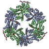
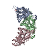
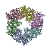
| Title | Crystal structure of the traffic ATPase (HP0525) of the Helicobacter pylori type IV secretion system bound by sulfate | ||||||
 Components Components | virB11 homolog | ||||||
 Keywords Keywords | HYDROLASE / VirB11 ATPase / bacterial type IV secretion / sulfate / HP0525 / Helicobacter pylori | ||||||
| Function / homology |  Function and homology information Function and homology informationsecretion by the type IV secretion system / type IV secretion system complex / nucleotide binding / ATP hydrolysis activity / metal ion binding / identical protein binding Similarity search - Function | ||||||
| Biological species |   Helicobacter pylori (bacteria) Helicobacter pylori (bacteria) | ||||||
| Method |  X-RAY DIFFRACTION / SYNCHROTRON / FOURIER SYNTHESIS / Resolution: 2.8 Å X-RAY DIFFRACTION / SYNCHROTRON / FOURIER SYNTHESIS / Resolution: 2.8 Å | ||||||
 Authors Authors | Savvides, S.N. / Yeo, H.J. / Beck, M.R. / Blaesing, F. / Lurz, R. / Lanka, E. / Buhrdorf, R. / Fischer, W. / Haas, R. / Waksman, G. | ||||||
 Citation Citation | Journal: Embo J. / Year: 2003 Title: VirB11 ATPases are dynamic hexameric assemblies: New insights into bacterial type IV secretion Authors: Savvides, S.N. / Yeo, H.J. / Beck, M.R. / Blaesing, F. / Lurz, R. / Lanka, E. / Buhrdorf, R. / Fischer, W. / Haas, R. / Waksman, G. | ||||||
| History |
|
- Structure visualization
| Structure viewer | Molecule: MolmilJmol/JSmol |
|---|
- Downloads & links
-Download
| PDBx/mmCIF format | 1opx.cif.gz | 138.7 KB | Display | PDBx/mmCIF format |
|---|---|---|---|---|
| PDB format | pdb1opx.ent.gz | 108.3 KB | Display | PDB format |
| PDBx/mmJSON format | 1opx.json.gz | Tree view | PDBx/mmJSON format | |
| Others |  Other downloads Other downloads |
-Validation report
| Arichive directory | https://data.pdbj.org/pub/pdb/validation_reports/op/1opxftp://data.pdbj.org/pub/pdb/validation_reports/op/1opx | HTTPS FTP |
|---|
-Related structure data


-Links
 PDBj
PDBj
- Assembly
| Deposited unit | 
| ||||||||
|---|---|---|---|---|---|---|---|---|---|
| 1 | 
| ||||||||
| 2 | x 6
| ||||||||
| Unit cell |
| ||||||||
| Details | The biologically relevant hecameric assembly is generated from the dimer in the asymmetric unit by a crystallographic three fold axis |
-Components
| #1: Protein | Mass: 37914.242 Da / Num. of mol.: 2 Source method: isolated from a genetically manipulated source Source: (gene. exp.) Helicobacter pylori (bacteria) / Strain: 26695 / Gene: hp0525 / Production host: #2: Chemical |   Mass: 96.063 Da / Num. of mol.: 2 / Source method: obtained synthetically / Formula: SO4 Mass: 96.063 Da / Num. of mol.: 2 / Source method: obtained synthetically / Formula: SO4#3: Chemical | ChemComp-2PE / |   Mass: 414.488 Da / Num. of mol.: 1 / Source method: obtained synthetically / Formula: C18H38O10 / Comment: precipitant*YM Mass: 414.488 Da / Num. of mol.: 1 / Source method: obtained synthetically / Formula: C18H38O10 / Comment: precipitant*YM#4: Water | ChemComp-HOH / |  Mass: 18.015 Da / Num. of mol.: 23 / Source method: isolated from a natural source / Formula: H2O Mass: 18.015 Da / Num. of mol.: 23 / Source method: isolated from a natural source / Formula: H2OHas protein modification | Y | |
|---|
-Experimental details
-Experiment
| Experiment | Method: X-RAY DIFFRACTION / Number of used crystals: 1 |
|---|
- Sample preparation
| Crystal | Density Matthews: 2.87 Å3/Da / Density % sol: 56.75 % | ||||||||||||||||||||||||
|---|---|---|---|---|---|---|---|---|---|---|---|---|---|---|---|---|---|---|---|---|---|---|---|---|---|
| Crystal grow | Temperature: 298 K / Method: vapor diffusion, hanging drop / pH: 7.8 Details: ammonium sulfate, peg 400, tris-HCl, pH 7.8, VAPOR DIFFUSION, HANGING DROP, temperature 298K | ||||||||||||||||||||||||
| Crystal grow | *PLUS Method: vapor diffusion | ||||||||||||||||||||||||
| Components of the solutions | *PLUS
|
-Data collection
| Diffraction | Mean temperature: 100 K |
|---|---|
| Diffraction source | Source: SYNCHROTRON / Site: APS  / Beamline: 19-BM / Wavelength: 0.9793 Å / Beamline: 19-BM / Wavelength: 0.9793 Å |
| Detector | Type: CUSTOM-MADE / Detector: CCD / Date: Sep 8, 2000 |
| Radiation | Protocol: SINGLE WAVELENGTH / Monochromatic (M) / Laue (L): M / Scattering type: x-ray |
| Radiation wavelength | Wavelength: 0.9793 Å / Relative weight: 1 |
| Reflection | Resolution: 2.8→30 Å / Num. all: 18699 / Num. obs: 18699 / % possible obs: 87 % / Observed criterion σ(F): 0 / Observed criterion σ(I): 0 / Redundancy: 4.2 % / Biso Wilson estimate: 11.3 Å2 / Rsym value: 0.115 |
| Reflection shell | Resolution: 2.8→2.89 Å / % possible all: 66 |
| Reflection | *PLUS Lowest resolution: 30 Å / Num. measured all: 80168 / Rmerge(I) obs: 0.115 |
| Reflection shell | *PLUS Lowest resolution: 2.9 Å / % possible obs: 94 % / Rmerge(I) obs: 0.156 / Mean I/σ(I) obs: 8.1 |
- Processing
| Software |
| ||||||||||||||||||||||||||||||||||||||||||||||||||||||||||||||||||||||||||||||||
|---|---|---|---|---|---|---|---|---|---|---|---|---|---|---|---|---|---|---|---|---|---|---|---|---|---|---|---|---|---|---|---|---|---|---|---|---|---|---|---|---|---|---|---|---|---|---|---|---|---|---|---|---|---|---|---|---|---|---|---|---|---|---|---|---|---|---|---|---|---|---|---|---|---|---|---|---|---|---|---|---|---|
| Refinement | Method to determine structure: FOURIER SYNTHESIS / Resolution: 2.8→29.63 Å / Rfactor Rfree error: 0.012 / Isotropic thermal model: RESTRAINED / Cross valid method: THROUGHOUT / σ(F): 2 / Stereochemistry target values: Engh & Huber
| ||||||||||||||||||||||||||||||||||||||||||||||||||||||||||||||||||||||||||||||||
| Solvent computation | Solvent model: FLAT MODEL / Bsol: 41.852 Å2 / ksol: 0.390312 e/Å3 | ||||||||||||||||||||||||||||||||||||||||||||||||||||||||||||||||||||||||||||||||
| Displacement parameters | Biso mean: 31.5 Å2
| ||||||||||||||||||||||||||||||||||||||||||||||||||||||||||||||||||||||||||||||||
| Refine analyze |
| ||||||||||||||||||||||||||||||||||||||||||||||||||||||||||||||||||||||||||||||||
| Refinement step | Cycle: LAST / Resolution: 2.8→29.63 Å
| ||||||||||||||||||||||||||||||||||||||||||||||||||||||||||||||||||||||||||||||||
| Refine LS restraints |
| ||||||||||||||||||||||||||||||||||||||||||||||||||||||||||||||||||||||||||||||||
| LS refinement shell | Resolution: 2.8→2.98 Å / Rfactor Rfree error: 0.04 / Total num. of bins used: 6
| ||||||||||||||||||||||||||||||||||||||||||||||||||||||||||||||||||||||||||||||||
| Xplor file |
| ||||||||||||||||||||||||||||||||||||||||||||||||||||||||||||||||||||||||||||||||
| Refinement | *PLUS Highest resolution: 2.8 Å / Lowest resolution: 30 Å / Rfactor Rwork: 0.25 | ||||||||||||||||||||||||||||||||||||||||||||||||||||||||||||||||||||||||||||||||
| Solvent computation | *PLUS | ||||||||||||||||||||||||||||||||||||||||||||||||||||||||||||||||||||||||||||||||
| Displacement parameters | *PLUS | ||||||||||||||||||||||||||||||||||||||||||||||||||||||||||||||||||||||||||||||||
| Refine LS restraints | *PLUS
|
