 Movie
Movie Controller

[English] 日本語
 Yorodumi
- PDB-2guk: Crystal Structure of the Conserved Protein of Unknown Function fr... -
+ Open data

- Basic information
| Entry | Database: PDB / ID: 2guk | ||||||
|---|---|---|---|---|---|---|---|
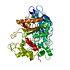
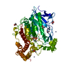
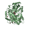
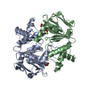
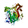
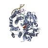
| Title | Crystal Structure of the Conserved Protein of Unknown Function from Porphyromonas gingivalis | ||||||
 Components Components | hypothetical protein PG1857 | ||||||
 Keywords Keywords | STRUCTURAL GENOMICS / UNKNOWN FUNCTION / alpha-beta / alpha-helical bundle / PSI / Protein Structure Initiative / Midwest Center for Structural Genomics / MCSG | ||||||
| Function / homology | PG1857-like / PG1857-like / Domain of unknown function DUF2023 / PG1857-like superfamily / Protein of unknown function (DUF2023) / 2-Layer Sandwich / Alpha Beta / : / DUF2023 domain-containing protein Function and homology information Function and homology information | ||||||
| Biological species |  Porphyromonas gingivalis (bacteria) Porphyromonas gingivalis (bacteria) | ||||||
| Method |  X-RAY DIFFRACTION / SYNCHROTRON / SAD / Resolution: 1.91 Å X-RAY DIFFRACTION / SYNCHROTRON / SAD / Resolution: 1.91 Å | ||||||
 Authors Authors | Kim, Y. / Quartey, P. / Moy, S. / Joachimiak, A. / Midwest Center for Structural Genomics (MCSG) | ||||||
 Citation Citation | Journal: To be Published Title: Crystal Structure of the Conserved Hypothetical Protein from Porphyromonas gingivalis Authors: Kim, Y. / Quartey, P. / Moy, S. / Joachimiak, A. | ||||||
| History |
| ||||||
| Remark 300 | THIS ENTRY CONTAINS THE CRYSTALLOGRAPHIC ASYMMETRIC UNIT WHICH CONSISTS OF 2 CHAIN(S). THERE IS NO ...THIS ENTRY CONTAINS THE CRYSTALLOGRAPHIC ASYMMETRIC UNIT WHICH CONSISTS OF 2 CHAIN(S). THERE IS NO EXPERIMENTAL EVIDENCE ABOUT THE BIOLOGICAL UNIT. THE BIOLOGICAL UNIT COULD BE A DIMER (AS IN THE ASYMMETRIC UNIT) OR A TETRAMER. SEE REMARK 350 FOR INFORMATION ON GENERATING THE BIOLOGICAL MOLECULE(S). |
- Structure visualization
| Structure viewer | Molecule: MolmilJmol/JSmol |
|---|
- Downloads & links
-Download
| PDBx/mmCIF format | 2guk.cif.gz | 62.9 KB | Display | PDBx/mmCIF format |
|---|---|---|---|---|
| PDB format | pdb2guk.ent.gz | 47.3 KB | Display | PDB format |
| PDBx/mmJSON format | 2guk.json.gz | Tree view | PDBx/mmJSON format | |
| Others |  Other downloads Other downloads |
-Validation report
| Arichive directory | https://data.pdbj.org/pub/pdb/validation_reports/gu/2gukftp://data.pdbj.org/pub/pdb/validation_reports/gu/2guk | HTTPS FTP |
|---|
-Related structure data
| Similar structure data | |
|---|---|
| Other databases |


-Links
 PDBj
PDBj- Assembly
| Deposited unit | 
| ||||||||
|---|---|---|---|---|---|---|---|---|---|
| 1 | 
| ||||||||
| Unit cell |
|
-Components
| #1: Protein | Mass: 14193.339 Da / Num. of mol.: 2 Source method: isolated from a genetically manipulated source Source: (gene. exp.) Porphyromonas gingivalis (bacteria) / Strain: W83 / Gene: PG1587, GeneID:2551814 / Plasmid: pMCSG7 / Species (production host): Escherichia coli / Production host: #2: Water | ChemComp-HOH / |  Mass: 18.015 Da / Num. of mol.: 132 / Source method: isolated from a natural source / Formula: H2O Mass: 18.015 Da / Num. of mol.: 132 / Source method: isolated from a natural source / Formula: H2OHas protein modification | Y | |
|---|
-Experimental details
-Experiment
| Experiment | Method: X-RAY DIFFRACTION / Number of used crystals: 1 |
|---|
- Sample preparation
| Crystal | Density Matthews: 2.06 Å3/Da / Density % sol: 40.27 % |
|---|---|
| Crystal grow | Temperature: 275 K / Method: vapor diffusion, sitting drop / pH: 7.5 Details: 5% iso-propanol, 0.1 M HEPES 7.5, 10 % PEG 4K, VAPOR DIFFUSION, SITTING DROP, temperature 275K |
-Data collection
| Diffraction | Mean temperature: 100 K |
|---|---|
| Diffraction source | Source: SYNCHROTRON / Site: APS  / Beamline: 19-ID / Wavelength: 0.97933 Å / Beamline: 19-ID / Wavelength: 0.97933 Å |
| Detector | Type: ADSC QUANTUM 315 / Detector: CCD / Date: Feb 24, 2006 / Details: mirrors |
| Radiation | Monochromator: double crystal monochromator, Si111 / Protocol: SINGLE WAVELENGTH / Monochromatic (M) / Laue (L): M / Scattering type: x-ray |
| Radiation wavelength | Wavelength: 0.97933 Å / Relative weight: 1 |
| Reflection | Resolution: 1.91→34.76 Å / Num. all: 20138 / Num. obs: 19279 / % possible obs: 98.6 % / Observed criterion σ(F): 0 / Observed criterion σ(I): 0 / Redundancy: 20.7 % / Rmerge(I) obs: 0.115 / Net I/σ(I): 10.5 |
| Reflection shell | Resolution: 1.91→1.98 Å / Redundancy: 11.4 % / Rmerge(I) obs: 0.82 / Mean I/σ(I) obs: 2.2 / Num. unique all: 1825 / % possible all: 96.7 |
- Processing
| Software |
| ||||||||||||||||||||||||||||||||||||||||||||||||||||||||||||||||||||||||||||||||||||||||||||||||||||||||||||||||||||||||||||||||||||||||||||||||||||||||||||||||||||||||||
|---|---|---|---|---|---|---|---|---|---|---|---|---|---|---|---|---|---|---|---|---|---|---|---|---|---|---|---|---|---|---|---|---|---|---|---|---|---|---|---|---|---|---|---|---|---|---|---|---|---|---|---|---|---|---|---|---|---|---|---|---|---|---|---|---|---|---|---|---|---|---|---|---|---|---|---|---|---|---|---|---|---|---|---|---|---|---|---|---|---|---|---|---|---|---|---|---|---|---|---|---|---|---|---|---|---|---|---|---|---|---|---|---|---|---|---|---|---|---|---|---|---|---|---|---|---|---|---|---|---|---|---|---|---|---|---|---|---|---|---|---|---|---|---|---|---|---|---|---|---|---|---|---|---|---|---|---|---|---|---|---|---|---|---|---|---|---|---|---|---|---|---|
| Refinement | Method to determine structure: SAD / Resolution: 1.91→34.76 Å / Cor.coef. Fo:Fc: 0.938 / Cor.coef. Fo:Fc free: 0.915 / SU B: 10.435 / SU ML: 0.153 / TLS residual ADP flag: LIKELY RESIDUAL / Cross valid method: THROUGHOUT / σ(F): 0 / σ(I): 0 / ESU R: 0.22 / ESU R Free: 0.192 Stereochemistry target values: MAXIMUM LIKELIHOOD WITH PHASES
| ||||||||||||||||||||||||||||||||||||||||||||||||||||||||||||||||||||||||||||||||||||||||||||||||||||||||||||||||||||||||||||||||||||||||||||||||||||||||||||||||||||||||||
| Solvent computation | Ion probe radii: 0.8 Å / Shrinkage radii: 0.8 Å / VDW probe radii: 1.4 Å / Solvent model: MASK | ||||||||||||||||||||||||||||||||||||||||||||||||||||||||||||||||||||||||||||||||||||||||||||||||||||||||||||||||||||||||||||||||||||||||||||||||||||||||||||||||||||||||||
| Displacement parameters | Biso mean: 31.333 Å2
| ||||||||||||||||||||||||||||||||||||||||||||||||||||||||||||||||||||||||||||||||||||||||||||||||||||||||||||||||||||||||||||||||||||||||||||||||||||||||||||||||||||||||||
| Refinement step | Cycle: LAST / Resolution: 1.91→34.76 Å
| ||||||||||||||||||||||||||||||||||||||||||||||||||||||||||||||||||||||||||||||||||||||||||||||||||||||||||||||||||||||||||||||||||||||||||||||||||||||||||||||||||||||||||
| Refine LS restraints |
| ||||||||||||||||||||||||||||||||||||||||||||||||||||||||||||||||||||||||||||||||||||||||||||||||||||||||||||||||||||||||||||||||||||||||||||||||||||||||||||||||||||||||||
| LS refinement shell | Resolution: 1.91→1.96 Å / Total num. of bins used: 20
| ||||||||||||||||||||||||||||||||||||||||||||||||||||||||||||||||||||||||||||||||||||||||||||||||||||||||||||||||||||||||||||||||||||||||||||||||||||||||||||||||||||||||||
| Refinement TLS params. | Method: refined / Refine-ID: X-RAY DIFFRACTION
| ||||||||||||||||||||||||||||||||||||||||||||||||||||||||||||||||||||||||||||||||||||||||||||||||||||||||||||||||||||||||||||||||||||||||||||||||||||||||||||||||||||||||||
| Refinement TLS group |
|
