 Movie
Movie Controller

[English] 日本語
 Yorodumi
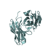
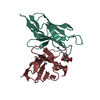
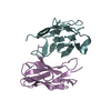
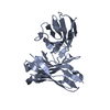
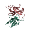
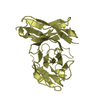
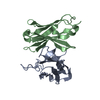
- PDB-2gsg: Crystal structure of the Fv fragment of a monoclonal antibody spe... -
+ Open data

- Basic information
| Entry | Database: PDB / ID: 2gsg | ||||||
|---|---|---|---|---|---|---|---|
| Title | Crystal structure of the Fv fragment of a monoclonal antibody specific for poly-glutamine | ||||||
 Components Components |
| ||||||
 Keywords Keywords | IMMUNE SYSTEM / Fv / monoclonal antibody / poly-glutamine | ||||||
| Function / homology | Immunoglobulins / Immunoglobulin-like / Sandwich / Mainly Beta Function and homology information Function and homology information | ||||||
| Biological species |  | ||||||
| Method |  X-RAY DIFFRACTION / SYNCHROTRON / MOLECULAR REPLACEMENT / Resolution: 2.1 Å X-RAY DIFFRACTION / SYNCHROTRON / MOLECULAR REPLACEMENT / Resolution: 2.1 Å | ||||||
 Authors Authors | Li, P. / Huey-Tubman, K.E. / West Jr., A.P. / Bennett, M.J. / Bjorkman, P.J. | ||||||
 Citation Citation | Journal: Nat.Struct.Mol.Biol. / Year: 2007 Title: The structure of a polyQ-anti-polyQ complex reveals binding according to a linear lattice model. Authors: Li, P. / Huey-Tubman, K.E. / Gao, T. / Li, X. / West Jr., A.P. / Bennett, M.J. / Bjorkman, P.J. | ||||||
| History |
|
- Structure visualization
| Structure viewer | Molecule: MolmilJmol/JSmol |
|---|
- Downloads & links
-Download
| PDBx/mmCIF format | 2gsg.cif.gz | 103.1 KB | Display | PDBx/mmCIF format |
|---|---|---|---|---|
| PDB format | pdb2gsg.ent.gz | 85.4 KB | Display | PDB format |
| PDBx/mmJSON format | 2gsg.json.gz | Tree view | PDBx/mmJSON format | |
| Others |  Other downloads Other downloads |
-Validation report
| Summary document | 2gsg_validation.pdf.gz | 448.4 KB | Display | wwPDB validaton report |
|---|---|---|---|---|
| Full document | 2gsg_full_validation.pdf.gz | 463.1 KB | Display | |
| Data in XML | 2gsg_validation.xml.gz | 23.4 KB | Display | |
| Data in CIF | 2gsg_validation.cif.gz | 33.1 KB | Display | |
| Arichive directory | https://data.pdbj.org/pub/pdb/validation_reports/gs/2gsgftp://data.pdbj.org/pub/pdb/validation_reports/gs/2gsg | HTTPS FTP |
-Related structure data
| Similar structure data |
|---|


-Links
 PDBj
PDBj

- Assembly
| Deposited unit | 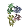
| ||||||||
|---|---|---|---|---|---|---|---|---|---|
| 1 | 
| ||||||||
| 2 | 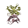
| ||||||||
| 3 | 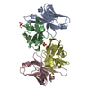
| ||||||||
| Unit cell |
|
-Components
| #1: Antibody | Mass: 12499.897 Da / Num. of mol.: 2 / Fragment: variable domain Source method: isolated from a genetically manipulated source Source: (gene. exp.)  #2: Antibody | Mass: 13330.936 Da / Num. of mol.: 2 / Fragment: variable domain Source method: isolated from a genetically manipulated source Source: (gene. exp.) #3: Chemical |   Mass: 96.063 Da / Num. of mol.: 3 / Source method: obtained synthetically / Formula: SO4 Mass: 96.063 Da / Num. of mol.: 3 / Source method: obtained synthetically / Formula: SO4#4: Water | ChemComp-HOH / |  Mass: 18.015 Da / Num. of mol.: 299 / Source method: isolated from a natural source / Formula: H2O Mass: 18.015 Da / Num. of mol.: 299 / Source method: isolated from a natural source / Formula: H2O |
|---|
-Experimental details
-Experiment
| Experiment | Method: X-RAY DIFFRACTION / Number of used crystals: 1 |
|---|
- Sample preparation
| Crystal | Density Matthews: 3.12 Å3/Da / Density % sol: 60.57 % |
|---|---|
| Crystal grow | Temperature: 290 K / Method: evaporation / pH: 6.5 Details: 0.1M sodium cacodylate, pH 6.5, 20% PEG 8000, 0.2M (NH4)2SO4, EVAPORATION, temperature 290K |
-Data collection
| Diffraction | Mean temperature: 100 K |
|---|---|
| Diffraction source | Source: SYNCHROTRON / Site: ALS  / Beamline: 8.2.2 / Wavelength: 0.9537 Å / Beamline: 8.2.2 / Wavelength: 0.9537 Å |
| Detector | Type: ADSC QUANTUM 315 / Detector: CCD / Date: Nov 5, 2004 |
| Radiation | Monochromator: Si 111 CHANNEL / Protocol: SINGLE WAVELENGTH / Monochromatic (M) / Laue (L): M / Scattering type: x-ray |
| Radiation wavelength | Wavelength: 0.9537 Å / Relative weight: 1 |
| Reflection | Resolution: 2.1→50 Å / Num. all: 38667 / Num. obs: 37635 / % possible obs: 96.6 % / Observed criterion σ(F): 0 / Observed criterion σ(I): 0 / Redundancy: 5.5 % / Rsym value: 0.068 / Net I/σ(I): 31.5 |
| Reflection shell | Resolution: 2.1→2.18 Å / Redundancy: 3.6 % / Rmerge(I) obs: 0.444 / Mean I/σ(I) obs: 3.2 / Num. unique all: 3303 / Rsym value: 0.444 / % possible all: 86.4 |
- Processing
| Software |
| ||||||||||||||||||||
|---|---|---|---|---|---|---|---|---|---|---|---|---|---|---|---|---|---|---|---|---|---|
| Refinement | Method to determine structure: MOLECULAR REPLACEMENT / Resolution: 2.1→50 Å / σ(F): 0 / σ(I): 0 / Stereochemistry target values: Engh & Huber
| ||||||||||||||||||||
| Refinement step | Cycle: LAST / Resolution: 2.1→50 Å
|
