[English] 日本語
Yorodumi
- PDB-2gsb: Solution structure of the second SH2 domain of human Ras GTPase-a... -

+
Open data


ID or keywords:

Loading...

-
Basic information

Entry
Database: PDB / ID: 2gsb
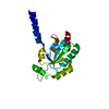
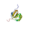
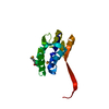
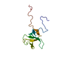
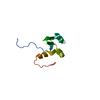
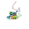
TitleSolution structure of the second SH2 domain of human Ras GTPase-activating protein 1
ComponentsRas GTPase-activating protein 1
KeywordsSIGNALING PROTEIN / GTPase-activating protein / GAP / Ras p21 protein activator / p120GAP / RasGAP / Structural Genomics / NPPSFA / National Project on Protein Structural and Functional Analyses / RIKEN Structural Genomics/Proteomics Initiative / RSGI
Function / homology
Function and homology information


regulation of RNA metabolic process / regulation of actin filament polymerization / potassium channel inhibitor activity / negative regulation of cell adhesion / blood vessel morphogenesis / negative regulation of cell-matrix adhesion / mitotic cytokinesis / ephrin receptor signaling pathway / vasculogenesis / PTK6 Regulates RHO GTPases, RAS GTPase and MAP kinases ...regulation of RNA metabolic process / regulation of actin filament polymerization / potassium channel inhibitor activity / negative regulation of cell adhesion / blood vessel morphogenesis / negative regulation of cell-matrix adhesion / mitotic cytokinesis / ephrin receptor signaling pathway / vasculogenesis / PTK6 Regulates RHO GTPases, RAS GTPase and MAP kinases / ruffle / EPHB-mediated forward signaling / phosphotyrosine residue binding / GTPase activator activity / Downstream signal transduction / VEGFR2 mediated cell proliferation / Regulation of RAS by GAPs / GTPase binding / regulation of cell shape / negative regulation of neuron apoptotic process / intracellular signal transduction / signaling receptor binding / GTPase activity / negative regulation of apoptotic process / signal transduction / plasma membrane / cytosol / cytoplasm
Similarity search - Function
Ras GTPase-activating protein 1, N-terminal SH2 domain / RasGAP, SH3 domain / Ras GTPase-activating protein 1, C-terminal SH2 domain / Ras GTPase-activating protein / Ras GTPase-activating protein, conserved site / Ras GTPase-activating proteins domain signature. / GTPase-activator protein for Ras-like GTPase / Ras GTPase-activating proteins profile. / GTPase-activator protein for Ras-like GTPases / Ras GTPase-activating domain ...Ras GTPase-activating protein 1, N-terminal SH2 domain / RasGAP, SH3 domain / Ras GTPase-activating protein 1, C-terminal SH2 domain / Ras GTPase-activating protein / Ras GTPase-activating protein, conserved site / Ras GTPase-activating proteins domain signature. / GTPase-activator protein for Ras-like GTPase / Ras GTPase-activating proteins profile. / GTPase-activator protein for Ras-like GTPases / Ras GTPase-activating domain / Rho GTPase activation protein / SH2 domain / C2 domain / Protein kinase C conserved region 2 (CalB) / SHC Adaptor Protein / C2 domain / C2 domain profile. / PH domain / C2 domain superfamily / PH domain profile. / Pleckstrin homology domain. / Pleckstrin homology domain / SH3 domain / SH2 domain / Src homology 2 (SH2) domain profile. / Src homology 2 domains / SH2 domain / Src homology 3 domains / SH2 domain superfamily / SH3-like domain superfamily / Src homology 3 (SH3) domain profile. / SH3 domain / PH-like domain superfamily / 2-Layer Sandwich / Alpha Beta
Similarity search - Domain/homology
Ras GTPase-activating protein 1
Similarity search - Component
Biological speciesHomo sapiens (human)
MethodSOLUTION NMR / torsion angle dynamics
AuthorsKurosaki, C. / Suetake, T. / Yoshida, M. / Hayashi, F. / Yokoyma, S. / RIKEN Structural Genomics/Proteomics Initiative (RSGI)
CitationJournal: To be Published
Title: Solution structure of the second SH2 domain of human Ras GTPase-activating protein 1
Authors: Kurosaki, C. / Suetake, T. / Yoshida, M. / Hayashi, F. / Yokoyma, S.
History
DepositionApr 26, 2006Deposition site: RCSB / Processing site: PDBJ
Revision 1.0May 1, 2007Provider: repository / Type: Initial release
Revision 1.1May 1, 2008Group: Version format compliance
Revision 1.2Jul 13, 2011Group: Version format compliance
Revision 1.3Mar 9, 2022Group: Data collection / Database references / Derived calculations
Category: database_2 / pdbx_nmr_software ...database_2 / pdbx_nmr_software / pdbx_struct_assembly / pdbx_struct_oper_list / struct_ref_seq_dif
Item: _database_2.pdbx_DOI / _database_2.pdbx_database_accession ..._database_2.pdbx_DOI / _database_2.pdbx_database_accession / _pdbx_nmr_software.name / _struct_ref_seq_dif.details

-
Structure visualization

Structure viewerMolecule:
MolmilJmol/JSmol

Downloads & links

-
Assembly

Deposited unit
A: Ras GTPase-activating protein 1


Theoretical massNumber of molelcules
Total (without water)13,5341
Polymers13,5341
Non-polymers00
Water0
1


  • Idetical with deposited unit
  • defined by author
TypeNameSymmetry operationNumber
identity operation1_555x,y,z1
NMR ensembles
DataCriteria
Number of conformers (submitted / calculated)20 / 100target function, structures with the lowest energy, structures with the least restraint violations
RepresentativeModel #1lowest energy

-
Components

#1: Protein Ras GTPase-activating protein 1 / GTPase-activating protein / GAP / Ras p21 protein activator / p120GAP / RasGAP


Mass: 13534.019 Da / Num. of mol.: 1 / Fragment: SH2 domain
Source method: isolated from a genetically manipulated source
Source: (gene. exp.) Homo sapiens (human) / Description: cell free protein synthesis / Gene: RASA1, RASA / Plasmid: P050711-17 / References: UniProt: P20936

-
Experimental details

-
Experiment

ExperimentMethod: SOLUTION NMR
NMR experiment
Conditions-IDExperiment-IDSolution-IDType
1113D 13C-separated NOESY
1213D 15N-separated NOESY

-
Sample preparation

DetailsContents: 1.27mM 13C, 15N-labeled protein; 20mM d-Tris-HCl (pH 7.0); 100mM NaCl; 1mM d-DTT; 0.02% NaN3, 10% D2O; 90% H2O
Solvent system: 10% D2O; 90% H2O
Sample conditionsIonic strength: 120 mM / pH: 7.0 / Pressure: ambient / Temperature: 293 K

-
NMR measurement

NMR spectrometerType: Varian INOVA / Manufacturer: Varian / Model: INOVA / Field strength: 800 MHz

-
Processing

NMR software
NameVersionDeveloperClassification
VNMR6.1cVariancollection
NMRPipe20031121Delaglio, F.processing
NMRView5.0.4Johnson, B. A.data analysis
KUJIRA0.934KOBAYASHI, N.data analysis
CYANA2.0.17Guntert, P.structure solution
CYANA2.0.17Guntert, P.refinement
RefinementMethod: torsion angle dynamics / Software ordinal: 1
NMR representativeSelection criteria: lowest energy
NMR ensembleConformer selection criteria: target function, structures with the lowest energy, structures with the least restraint violations
Conformers calculated total number: 100 / Conformers submitted total number: 20

+
About Yorodumi

-
News

-
Feb 9, 2022. New format data for meta-information of EMDB entries

New format data for meta-information of EMDB entries

  • Version 3 of the EMDB header file is now the official format.
  • The previous official version 1.9 will be removed from the archive.

Related info.:EMDB header

External links:wwPDB to switch to version 3 of the EMDB data model

-
Aug 12, 2020. Covid-19 info

Covid-19 info

URL: https://pdbj.org/emnavi/covid19.php

New page: Covid-19 featured information page in EM Navigator.

Related info.:Covid-19 info / Mar 5, 2020. Novel coronavirus structure data

+
Mar 5, 2020. Novel coronavirus structure data

Novel coronavirus structure data

Related info.:Yorodumi Speices / Aug 12, 2020. Covid-19 info

External links:COVID-19 featured content - PDBj / Molecule of the Month (242):Coronavirus Proteases

+
Jan 31, 2019. EMDB accession codes are about to change! (news from PDBe EMDB page)

EMDB accession codes are about to change! (news from PDBe EMDB page)

  • The allocation of 4 digits for EMDB accession codes will soon come to an end. Whilst these codes will remain in use, new EMDB accession codes will include an additional digit and will expand incrementally as the available range of codes is exhausted. The current 4-digit format prefixed with “EMD-” (i.e. EMD-XXXX) will advance to a 5-digit format (i.e. EMD-XXXXX), and so on. It is currently estimated that the 4-digit codes will be depleted around Spring 2019, at which point the 5-digit format will come into force.
  • The EM Navigator/Yorodumi systems omit the EMD- prefix.

Related info.:Q: What is EMD? / ID/Accession-code notation in Yorodumi/EM Navigator

External links:EMDB Accession Codes are Changing Soon! / Contact to PDBj

+
Jul 12, 2017. Major update of PDB

Major update of PDB

  • wwPDB released updated PDB data conforming to the new PDBx/mmCIF dictionary.
  • This is a major update changing the version number from 4 to 5, and with Remediation, in which all the entries are updated.
  • In this update, many items about electron microscopy experimental information are reorganized (e.g. em_software).
  • Now, EM Navigator and Yorodumi are based on the updated data.

External links:wwPDB Remediation / Enriched Model Files Conforming to OneDep Data Standards Now Available in the PDB FTP Archive

-
Yorodumi

Thousand views of thousand structures

  • Yorodumi is a browser for structure data from EMDB, PDB, SASBDB, etc.
  • This page is also the successor to EM Navigator detail page, and also detail information page/front-end page for Omokage search.
  • The word "yorodu" (or yorozu) is an old Japanese word meaning "ten thousand". "mi" (miru) is to see.

Related info.:EMDB / PDB / SASBDB / Comparison of 3 databanks / Yorodumi Search / Aug 31, 2016. New EM Navigator & Yorodumi / Yorodumi Papers / Jmol/JSmol / Function and homology information / Changes in new EM Navigator and Yorodumi

Read more