 Movie
Movie Controller

[English] 日本語
 Yorodumi
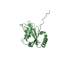
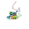
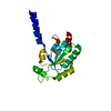
- PDB-2gm2: NMR structure of Xanthomonas campestris XCC1710: Northeast Struct... -
+ Open data

- Basic information
| Entry | Database: PDB / ID: 2gm2 | ||||||
|---|---|---|---|---|---|---|---|
| Title | NMR structure of Xanthomonas campestris XCC1710: Northeast Structural Genomics Consortium target XcR35 | ||||||
 Components Components | conserved hypothetical protein | ||||||
 Keywords Keywords | STRUCTURAL GENOMICS / UNKNOWN FUNCTION / MTH938-like fold / PSI / Protein Structure Initiative / Northeast Structural Genomics Consortium / NESG | ||||||
| Function / homology | Hypothetical Protein Mth938; Chain: A, / MTH938-like / NDUFAF3/Mth938 domain-containing protein / MTH938-like superfamily / Protein of unknown function (DUF498/DUF598) / 3-Layer(aba) Sandwich / Alpha Beta / : / Xcc1710-like domain-containing protein Function and homology information Function and homology information | ||||||
| Biological species |  Xanthomonas campestris pv. campestris str. ATCC 33913 (bacteria) Xanthomonas campestris pv. campestris str. ATCC 33913 (bacteria) | ||||||
| Method | SOLUTION NMR / THE INITIAL STRUCTURE WAS DETERMINED USING AUTOMATED STRUCTURE DETERMINATION (AUTOSTRUCTURE), REFINED MANUALLY. A FINAL REFINEMENT USED SIMULTATED ANNEALING IN EXPLICIT SOLVENT. | ||||||
 Authors Authors | Cort, J.R. / Xiao, R. / Wang, D.Y. / Ma, L.C. / Ciano, M. / Montelione, G.T. / Ramelot, T.A. / Kennedy, M.A. / Northeast Structural Genomics Consortium (NESG) | ||||||
 Citation Citation | Journal: To be Published Title: NMR structure of Xanthomonas campestris XCC1710 protein Authors: Cort, J.R. / Ramelot, T.A. / Montelione, G.T. / Kennedy, M.A. | ||||||
| History |
|
- Structure visualization
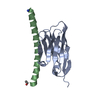
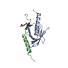
| Structure viewer | Molecule: MolmilJmol/JSmol |
|---|
- Downloads & links
-Download
| PDBx/mmCIF format | 2gm2.cif.gz | 787.2 KB | Display | PDBx/mmCIF format |
|---|---|---|---|---|
| PDB format | pdb2gm2.ent.gz | 659.1 KB | Display | PDB format |
| PDBx/mmJSON format | 2gm2.json.gz | Tree view | PDBx/mmJSON format | |
| Others |  Other downloads Other downloads |
-Validation report
| Arichive directory | https://data.pdbj.org/pub/pdb/validation_reports/gm/2gm2ftp://data.pdbj.org/pub/pdb/validation_reports/gm/2gm2 | HTTPS FTP |
|---|
-Related structure data
| Related structure data | |
|---|---|
| Similar structure data | |
| Other databases |


-Links
 PDBj
PDBj- Assembly
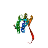
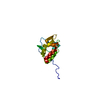
| Deposited unit | 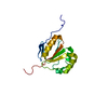
| |||||||||
|---|---|---|---|---|---|---|---|---|---|---|
| 1 |
| |||||||||
| NMR ensembles |
|
-Components
| #1: Protein | Mass: 14468.479 Da / Num. of mol.: 1 Source method: isolated from a genetically manipulated source Source: (gene. exp.) Xanthomonas campestris pv. campestris str. ATCC 33913 (bacteria)Species: Xanthomonas campestris / Strain: pv. campestris str. ATCC 33913 / Gene: XCC1710 / Plasmid: pET21 / Production host: |
|---|
-Experimental details
-Experiment
| Experiment | Method: SOLUTION NMR | ||||||||||||||||||||||||
|---|---|---|---|---|---|---|---|---|---|---|---|---|---|---|---|---|---|---|---|---|---|---|---|---|---|
| NMR experiment |
|
- Sample preparation
| Details |
| |||||||||
|---|---|---|---|---|---|---|---|---|---|---|
| Sample conditions | Ionic strength: 100 mM NaCl, 20 mM CaCl2 / pH: 6.5 / Pressure: ambient / Temperature: 298 K |
-NMR measurement
| Radiation | Protocol: SINGLE WAVELENGTH / Monochromatic (M) / Laue (L): M | ||||||||||||||||||||
|---|---|---|---|---|---|---|---|---|---|---|---|---|---|---|---|---|---|---|---|---|---|
| Radiation wavelength | Relative weight: 1 | ||||||||||||||||||||
| NMR spectrometer |
|
- Processing
| NMR software |
| ||||||||||||||||||||||||||||||||
|---|---|---|---|---|---|---|---|---|---|---|---|---|---|---|---|---|---|---|---|---|---|---|---|---|---|---|---|---|---|---|---|---|---|
| Refinement | Method: THE INITIAL STRUCTURE WAS DETERMINED USING AUTOMATED STRUCTURE DETERMINATION (AUTOSTRUCTURE), REFINED MANUALLY. A FINAL REFINEMENT USED SIMULTATED ANNEALING IN EXPLICIT SOLVENT. Software ordinal: 1 Details: THE STRUCTURES ARE BASED ON A TOTAL OF 935 RESTRAINTS. SUMMARY OF EXPERIMENTAL CONSTRAINTS RESTRAINING DISTANCE RESTRAINTS: TOTAL = 758; INTRA-RESIDUE [I=J] = 174; SEQUENTIAL [(I-J)=1] = 174; ...Details: THE STRUCTURES ARE BASED ON A TOTAL OF 935 RESTRAINTS. SUMMARY OF EXPERIMENTAL CONSTRAINTS RESTRAINING DISTANCE RESTRAINTS: TOTAL = 758; INTRA-RESIDUE [I=J] = 174; SEQUENTIAL [(I-J)=1] = 174; MEDIUM RANGE [1<(I-J)<5] = 109; LONG RANGE [(I-J)>=5] = 301; HYDROGEN BOND RESTRAINTS = 56 (2 PER H-BOND); NUMBER OF RESTRAINING DISTANCE RESTRAINTS PER RESTRAINED RESIDUE = 7.3; DIHEDRAL-ANGLE RESTRAINTS = 121 (60 PHI, 59 PSI, 2 CHI-1); TOTAL NUMBER OF RESTRAINTS PER RESTRAINED RESIDUE = 8.3; NUMBER OF LONG RANGE NOE DISTANCE RESTRAINTS PER RESTRAINED RESIDUE = 2.7; NUMBER OF STRUCTURES COMPUTED = 40; NUMBER OF STRUCTURES USED = 20; AVERAGE DISTANCE VIOLATIONS >0.0001 ANG = 19.8 +/- 3.5; AVERAGE R.M.S. DISTANCE VIOLATION = 0.0009 +/- 0.0003 ANG; MAXIMUM NUMBER OF DISTANCE VIOLATIONS 25; MAXIMUM DISTANCE VIOLATION = 0.03 ANG; AVERAGE DIHEDRAL ANGLE VIOLATIONS: >0.0001 DEG = 2.5+/-1.3; MAX NUMBER OF DIHEDRAL ANGLE VIOLATIONS = 4; AVERAGE R.M.S. DIHEDRAL ANGLE VIOLATION = 0.02 +/- .01 DEG.; RMSD VALUES TO AVERAGE STRUCTURE: BACKBONE ATOMS (N,C,C' RESIDUES 12-125) = 0.88 ANG, ALL HEAVY ATOMS = 1.38 ANG; BACKBONE ATOMS (N,C,C' RESIDUES 32-122) = 0.71 ANG, ALL HEAVY ATOMS = 1.21 ANG; BACKBONE ATOMS (N,C,C' RESIDUES 16-17,21-36,39-41,44-46,53-73,76-122) = 0.70 ANG, ALL HEAVY ATOMS = 1.14 ANG; PROCHECK (RESIDUES 16-17,21-36,39-41,44-46,53-73,76-122): MOST FAVORED REGIONS = 84.6%; ADDITIONAL ALLOWED REGIONS = 14.0%; GENEROUSLY ALLOWED REGIONS = 0.2%; DISALLOWED REGIONS = 1.2%; PROCHECK (RESIDUES 12-125): MOST FAVORED REGIONS = 77.8%; ADDITIONAL ALLOWED REGIONS = 19.2%; GENEROUSLY ALLOWED REGIONS = 2.0%; DISALLOWED REGIONS = 1.0%. | ||||||||||||||||||||||||||||||||
| NMR representative | Selection criteria: closest to the average | ||||||||||||||||||||||||||||||||
| NMR ensemble | Conformer selection criteria: structures with fewest restraint violations, low restraint violation energies, and acceptable geometry Conformers calculated total number: 40 / Conformers submitted total number: 20 |
 X-PLOR