 Movie
Movie Controller


+ Open data

- Basic information
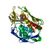
| Entry | Database: PDB / ID: 2go4 | ||||||
|---|---|---|---|---|---|---|---|
| Title | Crystal structure of Aquifex aeolicus LpxC complexed with TU-514 | ||||||
 Components Components | UDP-3-O-[3-hydroxymyristoyl] N-acetylglucosamine deacetylase | ||||||
 Keywords Keywords | HYDROLASE / LpxC-inhibitor complex | ||||||
| Function / homology |  Function and homology information Function and homology informationUDP-3-O-acyl-N-acetylglucosamine deacetylase / UDP-3-O-acyl-N-acetylglucosamine deacetylase activity / lipid A biosynthetic process / membrane / metal ion binding / cytoplasm Similarity search - Function | ||||||
| Biological species |   Aquifex aeolicus (bacteria) Aquifex aeolicus (bacteria) | ||||||
| Method |  X-RAY DIFFRACTION / SYNCHROTRON / FOURIER SYNTHESIS / Resolution: 2.7 Å X-RAY DIFFRACTION / SYNCHROTRON / FOURIER SYNTHESIS / Resolution: 2.7 Å | ||||||
 Authors Authors | Gennadios, H.A. / Whittington, D.A. / Li, X. / Fierke, C.A. / Christianson, D.W. | ||||||
 Citation Citation | Journal: Biochemistry / Year: 2006 Title: Mechanistic Inferences from the Binding of Ligands to LpxC, a Metal-Dependent Deacetylase Authors: Gennadios, H.A. / Whittington, D.A. / Li, X. / Fierke, C.A. / Christianson, D.W. | ||||||
| History |
| ||||||
| Remark 300 | BIOMOLECULE: 1, 2 THIS ENTRY CONTAINS THE CRYSTALLOGRAPHIC ASYMMETRIC UNIT WHICH CONSISTS OF 2 ...BIOMOLECULE: 1, 2 THIS ENTRY CONTAINS THE CRYSTALLOGRAPHIC ASYMMETRIC UNIT WHICH CONSISTS OF 2 CHAIN(S). ALTHOUGH IN THE CRYSTAL THIS MOLECULE APPEARS TO FORM A DIMER, THE MONOMER IS BIOLOGICALLY ACTIVE. SEE REMARK 350 FOR INFORMATION ON GENERATING THE BIOLOGICAL MOLECULE(S). | ||||||
| Remark 999 | SEQUENCE THE RESIDUE NUMBERING SCHEME FOR THIS STRUCTURE FOLLOWS THAT OF THE E. COLI ENZYME. THIS ...SEQUENCE THE RESIDUE NUMBERING SCHEME FOR THIS STRUCTURE FOLLOWS THAT OF THE E. COLI ENZYME. THIS TREATMENT RESULTS IN A BREAK IN THE SEQUENTIAL NUMBERING IN A COUPLE OF PLACES. |
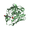
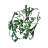
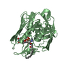
- Structure visualization
| Structure viewer | Molecule: MolmilJmol/JSmol |
|---|
- Downloads & links
-Download
| PDBx/mmCIF format | 2go4.cif.gz | 123.1 KB | Display | PDBx/mmCIF format |
|---|---|---|---|---|
| PDB format | pdb2go4.ent.gz | 95 KB | Display | PDB format |
| PDBx/mmJSON format | 2go4.json.gz | Tree view | PDBx/mmJSON format | |
| Others |  Other downloads Other downloads |
-Validation report
| Arichive directory | https://data.pdbj.org/pub/pdb/validation_reports/go/2go4ftp://data.pdbj.org/pub/pdb/validation_reports/go/2go4 | HTTPS FTP |
|---|
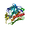
-Related structure data
| Related structure data | 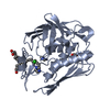 2go3C  1p42S S: Starting model for refinement C: citing same article ( |
|---|---|
| Similar structure data |


-Links
 PDBj
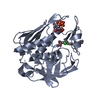
PDBj- Assembly
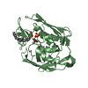
| Deposited unit | 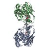
| ||||||||
|---|---|---|---|---|---|---|---|---|---|
| 1 | 
| ||||||||
| 2 | 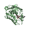
| ||||||||
| 3 | 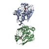
| ||||||||
| Unit cell |
| ||||||||
| Details | Biological monomer, however, crystallographic dimer |
-Components
| #1: Protein | Mass: 30488.875 Da / Num. of mol.: 2 / Fragment: delta 11 C-terminal deletion / Mutation: C193A Source method: isolated from a genetically manipulated source Source: (gene. exp.) Aquifex aeolicus (bacteria) / Gene: lpxC, envA / Plasmid: pET21a / Production host: References: UniProt: O67648, Hydrolases; Acting on carbon-nitrogen bonds, other than peptide bonds; In linear amides #2: Chemical | ChemComp-ZN /   Mass: 65.409 Da / Num. of mol.: 5 / Source method: obtained synthetically / Formula: Zn Mass: 65.409 Da / Num. of mol.: 5 / Source method: obtained synthetically / Formula: Zn#3: Chemical | ChemComp-CL / |   Mass: 35.453 Da / Num. of mol.: 1 / Source method: obtained synthetically / Formula: Cl Mass: 35.453 Da / Num. of mol.: 1 / Source method: obtained synthetically / Formula: Cl#4: Chemical | 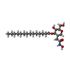  Mass: 431.563 Da / Num. of mol.: 2 / Source method: obtained synthetically / Formula: C22H41NO7 Mass: 431.563 Da / Num. of mol.: 2 / Source method: obtained synthetically / Formula: C22H41NO7#5: Water | ChemComp-HOH / |  Mass: 18.015 Da / Num. of mol.: 56 / Source method: isolated from a natural source / Formula: H2O Mass: 18.015 Da / Num. of mol.: 56 / Source method: isolated from a natural source / Formula: H2O |
|---|
-Experimental details
-Experiment
| Experiment | Method: X-RAY DIFFRACTION / Number of used crystals: 1 |
|---|
- Sample preparation
| Crystal | Density Matthews: 2.91 Å3/Da / Density % sol: 57.77 % |
|---|---|
| Crystal grow | Temperature: 294 K / Method: vapor diffusion, hanging drop / pH: 7.5 Details: 100 mM HEPES (pH 7.5), 180 mM NaCl, 14% PEG 3350, 5 mM ZnSO4, VAPOR DIFFUSION, HANGING DROP, temperature 294K |
-Data collection
| Diffraction | Mean temperature: 200 K |
|---|---|
| Diffraction source | Source: SYNCHROTRON / Site: CHESS  / Beamline: F1 / Wavelength: 0.9124 Å / Beamline: F1 / Wavelength: 0.9124 Å |
| Detector | Type: ADSC QUANTUM 4 / Detector: CCD / Date: Feb 10, 2006 / Details: Rh coated Si monochromatic mirror |
| Radiation | Monochromator: Horizontal bent Si(111), asymmetrically cut with water cooled Cu Block Protocol: SINGLE WAVELENGTH / Monochromatic (M) / Laue (L): M / Scattering type: x-ray |
| Radiation wavelength | Wavelength: 0.9124 Å / Relative weight: 1 |
| Reflection | Resolution: 2.7→50 Å / Num. all: 19244 / Num. obs: 17601 / % possible obs: 97.7 % / Rmerge(I) obs: 0.069 / Net I/σ(I): 12.6 |
| Reflection shell | Resolution: 2.7→2.8 Å / Mean I/σ(I) obs: 2.2 / % possible all: 97.2 |
- Processing
| Software |
| ||||||||||||||||||||||||||||
|---|---|---|---|---|---|---|---|---|---|---|---|---|---|---|---|---|---|---|---|---|---|---|---|---|---|---|---|---|---|
| Refinement | Method to determine structure: FOURIER SYNTHESIS Starting model: 1P42 (zinc-inhibited LpxC) Resolution: 2.7→50 Å / Stereochemistry target values: Engh & Huber
| ||||||||||||||||||||||||||||
| Refine analyze | Luzzati coordinate error obs: 0.33 Å / Luzzati d res low obs: 5 Å / Luzzati sigma a obs: 0.39 Å | ||||||||||||||||||||||||||||
| Refinement step | Cycle: LAST / Resolution: 2.7→50 Å
| ||||||||||||||||||||||||||||
| Refine LS restraints |
| ||||||||||||||||||||||||||||
| LS refinement shell |
|
