 Movie
Movie Controller

[English] 日本語
 Yorodumi
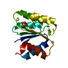
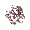
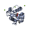
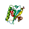
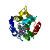
- PDB-2gkg: Receiver domain from Myxococcus xanthus social motility protein FrzS -
+ Open data

- Basic information
| Entry | Database: PDB / ID: 2gkg | ||||||
|---|---|---|---|---|---|---|---|
| Title | Receiver domain from Myxococcus xanthus social motility protein FrzS | ||||||
 Components Components | response regulator homolog | ||||||
 Keywords Keywords | SIGNALING PROTEIN / social motility / receiver domain / signalling / high resolution | ||||||
| Function / homology |  Function and homology information Function and homology information | ||||||
| Biological species |  Myxococcus xanthus (bacteria) Myxococcus xanthus (bacteria) | ||||||
| Method |  X-RAY DIFFRACTION / SYNCHROTRON / MOLECULAR REPLACEMENT / Resolution: 1 Å X-RAY DIFFRACTION / SYNCHROTRON / MOLECULAR REPLACEMENT / Resolution: 1 Å | ||||||
 Authors Authors | Echols, N. / Fraser, J. / Merlie, J. / Zusman, D. / Alber, T. | ||||||
 Citation Citation | Journal: Mol.Microbiol. / Year: 2007 Title: An atypical receiver domain controls the dynamic polar localization of the Myxococcus xanthus social motility protein FrzS. Authors: Fraser, J.S. / Merlie, J.P. / Echols, N. / Weisfield, S.R. / Mignot, T. / Wemmer, D.E. / Zusman, D.R. / Alber, T. | ||||||
| History |
|
- Structure visualization
| Structure viewer | Molecule: MolmilJmol/JSmol |
|---|
- Downloads & links
-Download
| PDBx/mmCIF format | 2gkg.cif.gz | 85.3 KB | Display | PDBx/mmCIF format |
|---|---|---|---|---|
| PDB format | pdb2gkg.ent.gz | 63.9 KB | Display | PDB format |
| PDBx/mmJSON format | 2gkg.json.gz | Tree view | PDBx/mmJSON format | |
| Others |  Other downloads Other downloads |
-Validation report
| Arichive directory | https://data.pdbj.org/pub/pdb/validation_reports/gk/2gkgftp://data.pdbj.org/pub/pdb/validation_reports/gk/2gkg | HTTPS FTP |
|---|
-Related structure data
| Related structure data | 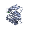 2i6fC  2nt3C  2nt4C  1w25S C: citing same article ( S: Starting model for refinement |
|---|---|
| Similar structure data |


-Links
 PDBj
PDBj

- Assembly
| Deposited unit | 
| ||||||||
|---|---|---|---|---|---|---|---|---|---|
| 1 |
| ||||||||
| Unit cell |
| ||||||||
| Details | ASU contains one copy of FrzS receiver domain. The full-length protein includes a coiled-coil domain and may oligomerize. |
-Components
| #1: Protein | Mass: 13686.523 Da / Num. of mol.: 1 / Fragment: receiver domain (residues 1-124) Source method: isolated from a genetically manipulated source Source: (gene. exp.) Myxococcus xanthus (bacteria) / Gene: FrzS / Plasmid: pET28b / Species (production host): Escherichia coli / Production host: |
|---|---|
| #2: Water | ChemComp-HOH /  Mass: 18.015 Da / Num. of mol.: 361 / Source method: isolated from a natural source / Formula: H2O Mass: 18.015 Da / Num. of mol.: 361 / Source method: isolated from a natural source / Formula: H2O |
-Experimental details
-Experiment
| Experiment | Method: X-RAY DIFFRACTION / Number of used crystals: 1 |
|---|
- Sample preparation
| Crystal | Density Matthews: 2.01 Å3/Da / Density % sol: 38.82 % |
|---|---|
| Crystal grow | Temperature: 291 K / Method: vapor diffusion, hanging drop Details: PEG 3350, NaFormate, VAPOR DIFFUSION, HANGING DROP, temperature 291K |
-Data collection
| Diffraction | Mean temperature: 100 K |
|---|---|
| Diffraction source | Source: SYNCHROTRON / Site: ALS  / Beamline: 8.3.1 / Wavelength: 0.8856 / Wavelength: 0.8856 Å / Beamline: 8.3.1 / Wavelength: 0.8856 / Wavelength: 0.8856 Å |
| Detector | Type: ADSC QUANTUM 210 / Detector: CCD / Date: Nov 9, 2005 |
| Radiation | Monochromator: SI 111 / Protocol: SINGLE WAVELENGTH / Monochromatic (M) / Laue (L): M / Scattering type: x-ray |
| Radiation wavelength | Wavelength: 0.8856 Å / Relative weight: 1 |
| Reflection | Resolution: 1→20 Å / Num. all: 58567 / Num. obs: 58567 / % possible obs: 99.98 % / Observed criterion σ(F): 0 / Observed criterion σ(I): 0 / Redundancy: 8.5 % / Rmerge(I) obs: 0.061 / Net I/σ(I): 28.75 |
| Reflection shell | Resolution: 1→1.02 Å / Redundancy: 6.3 % / Rmerge(I) obs: 0.492 / Mean I/σ(I) obs: 2 / % possible all: 100 |
- Processing
| Software |
| |||||||||||||||||||||||||||||||||
|---|---|---|---|---|---|---|---|---|---|---|---|---|---|---|---|---|---|---|---|---|---|---|---|---|---|---|---|---|---|---|---|---|---|---|
| Refinement | Method to determine structure: MOLECULAR REPLACEMENT Starting model: GUANYLATE CYCLASE RECEIVER DOMAIN (PDB CODE: 1W25) Resolution: 1→20 Å / Num. parameters: 13613 / Num. restraintsaints: 18431 / Cross valid method: FREE R / σ(F): 0 / Stereochemistry target values: ENGH AND HUBER Details: All hydrogen atoms on protein residues were included in the final round of refinement. Positions of hydroxyl and methyl hydrogens were refined independently.
| |||||||||||||||||||||||||||||||||
| Refine analyze | Num. disordered residues: 31 / Occupancy sum hydrogen: 947 / Occupancy sum non hydrogen: 1204.95 | |||||||||||||||||||||||||||||||||
| Refinement step | Cycle: LAST / Resolution: 1→20 Å
| |||||||||||||||||||||||||||||||||
| Refine LS restraints |
|
