 Movie
Movie Controller

[English] 日本語
 Yorodumi
- PDB-2gib: Crystal structure of the SARS coronavirus nucleocapsid protein di... -
+ Open data

- Basic information
| Entry | Database: PDB / ID: 2gib | ||||||
|---|---|---|---|---|---|---|---|
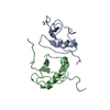
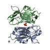
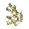
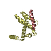
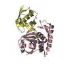
| Title | Crystal structure of the SARS coronavirus nucleocapsid protein dimerization domain | ||||||
 Components Components | Nucleocapsid protein | ||||||
 Keywords Keywords | VIRAL PROTEIN / SARS / nucleocapsid / dimer | ||||||
| Function / homology |  Function and homology information Function and homology informationSARS-CoV-1-host interactions / Translation of Structural Proteins / Virion Assembly and Release / viral RNA genome packaging / Transcription of SARS-CoV-1 sgRNAs / negative regulation of interferon-beta production / Maturation of nucleoprotein / SARS-CoV-1 modulates host translation machinery / SARS-CoV-1 targets host intracellular signalling and regulatory pathways / Attachment and Entry ...SARS-CoV-1-host interactions / Translation of Structural Proteins / Virion Assembly and Release / viral RNA genome packaging / Transcription of SARS-CoV-1 sgRNAs / negative regulation of interferon-beta production / Maturation of nucleoprotein / SARS-CoV-1 modulates host translation machinery / SARS-CoV-1 targets host intracellular signalling and regulatory pathways / Attachment and Entry / SARS-CoV-1 activates/modulates innate immune responses / viral capsid / viral nucleocapsid / host cell endoplasmic reticulum-Golgi intermediate compartment / molecular adaptor activity / host cell Golgi apparatus / host cell perinuclear region of cytoplasm / ribonucleoprotein complex / host cell nucleus / DNA binding / RNA binding / identical protein binding / plasma membrane Similarity search - Function | ||||||
| Biological species |  SARS coronavirus SARS coronavirus | ||||||
| Method |  X-RAY DIFFRACTION / SYNCHROTRON / MAD / Resolution: 1.75 Å X-RAY DIFFRACTION / SYNCHROTRON / MAD / Resolution: 1.75 Å | ||||||
 Authors Authors | Yu, I.M. / Oldham, M.L. / Zhang, J. / Chen, J. | ||||||
 Citation Citation | Journal: J.Biol.Chem. / Year: 2006 Title: Crystal structure of the severe acute respiratory syndrome (SARS) coronavirus nucleocapsid protein dimerization domain reveals evolutionary linkage between corona- and arteriviridae. Authors: Yu, I.M. / Oldham, M.L. / Zhang, J. / Chen, J. | ||||||
| History |
|
- Structure visualization
| Structure viewer | Molecule: MolmilJmol/JSmol |
|---|
- Downloads & links
-Download
| PDBx/mmCIF format | 2gib.cif.gz | 94.2 KB | Display | PDBx/mmCIF format |
|---|---|---|---|---|
| PDB format | pdb2gib.ent.gz | 72.8 KB | Display | PDB format |
| PDBx/mmJSON format | 2gib.json.gz | Tree view | PDBx/mmJSON format | |
| Others |  Other downloads Other downloads |
-Validation report
| Arichive directory | https://data.pdbj.org/pub/pdb/validation_reports/gi/2gibftp://data.pdbj.org/pub/pdb/validation_reports/gi/2gib | HTTPS FTP |
|---|
-Related structure data
| Similar structure data |
|---|


-Links
 PDBj
PDBj


- Assembly
| Deposited unit | 
| |||||||||
|---|---|---|---|---|---|---|---|---|---|---|
| 1 |
| |||||||||
| 2 | 
| |||||||||
| 3 | 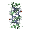
| |||||||||
| Unit cell |
| |||||||||
| Components on special symmetry positions |
| |||||||||
| Details | The biologic assembly is a dimer as found in the asymmetric unit |
-Components
| #1: Protein | Mass: 11554.877 Da / Num. of mol.: 2 / Fragment: dimerization domain (residues 270-370) Source method: isolated from a genetically manipulated source Source: (gene. exp.) SARS coronavirus / Genus: Coronavirus / Gene: N / Plasmid: pMCSG7 / Production host:  #2: Chemical | ChemComp-SO4 / |   Mass: 96.063 Da / Num. of mol.: 1 / Source method: obtained synthetically / Formula: SO4 Mass: 96.063 Da / Num. of mol.: 1 / Source method: obtained synthetically / Formula: SO4#3: Water | ChemComp-HOH / |  Mass: 18.015 Da / Num. of mol.: 212 / Source method: isolated from a natural source / Formula: H2O Mass: 18.015 Da / Num. of mol.: 212 / Source method: isolated from a natural source / Formula: H2O |
|---|
-Experimental details
-Experiment
| Experiment | Method: X-RAY DIFFRACTION / Number of used crystals: 1 |
|---|
- Sample preparation
| Crystal | Density Matthews: 2.7 Å3/Da / Density % sol: 54.38 % |
|---|---|
| Crystal grow | Temperature: 277.5 K / Method: vapor diffusion, sitting drop / pH: 6.5 Details: 30-33% Pentaerythritol ethoxylate 15/4 EO/OH, 50 mM Ammonium sulfate, 50 mM Bis-Tris , pH 6.5, VAPOR DIFFUSION, SITTING DROP, temperature 277.5K |
-Data collection
| Diffraction | Mean temperature: 100 K | ||||||||||||
|---|---|---|---|---|---|---|---|---|---|---|---|---|---|
| Diffraction source | Source: SYNCHROTRON / Site: APS  / Beamline: 19-ID / Wavelength: 0.97929, 0.97940, 0.94285 / Beamline: 19-ID / Wavelength: 0.97929, 0.97940, 0.94285 | ||||||||||||
| Detector | Type: ADSC QUANTUM 315 / Detector: CCD / Date: Nov 25, 2005 | ||||||||||||
| Radiation | Monochromator: Rosenbaum-Rock monochromator / Protocol: MAD / Monochromatic (M) / Laue (L): M / Scattering type: x-ray | ||||||||||||
| Radiation wavelength |
| ||||||||||||
| Reflection | Resolution: 1.75→30.93 Å / Num. all: 23293 / Num. obs: 23088 / % possible obs: 99.12 % / Observed criterion σ(F): 0 / Observed criterion σ(I): 0 | ||||||||||||
| Reflection shell | Resolution: 1.75→1.795 Å / % possible all: 96.12 |
- Processing
| Software |
| |||||||||||||||||||||||||||||||||||||||||||||||||||||||||||||||||||||||||||||||||||||||||||||||||||||||||
|---|---|---|---|---|---|---|---|---|---|---|---|---|---|---|---|---|---|---|---|---|---|---|---|---|---|---|---|---|---|---|---|---|---|---|---|---|---|---|---|---|---|---|---|---|---|---|---|---|---|---|---|---|---|---|---|---|---|---|---|---|---|---|---|---|---|---|---|---|---|---|---|---|---|---|---|---|---|---|---|---|---|---|---|---|---|---|---|---|---|---|---|---|---|---|---|---|---|---|---|---|---|---|---|---|---|---|
| Refinement | Method to determine structure: MAD / Resolution: 1.75→30.93 Å / Cor.coef. Fo:Fc: 0.938 / Cor.coef. Fo:Fc free: 0.914 / SU B: 4.535 / SU ML: 0.069 / Cross valid method: THROUGHOUT / σ(F): 0 / ESU R: 0.16 / ESU R Free: 0.114 / Stereochemistry target values: MAXIMUM LIKELIHOOD
| |||||||||||||||||||||||||||||||||||||||||||||||||||||||||||||||||||||||||||||||||||||||||||||||||||||||||
| Solvent computation | Ion probe radii: 0.8 Å / Shrinkage radii: 0.8 Å / VDW probe radii: 1.2 Å / Solvent model: MASK | |||||||||||||||||||||||||||||||||||||||||||||||||||||||||||||||||||||||||||||||||||||||||||||||||||||||||
| Displacement parameters | Biso mean: 17.965 Å2
| |||||||||||||||||||||||||||||||||||||||||||||||||||||||||||||||||||||||||||||||||||||||||||||||||||||||||
| Refinement step | Cycle: LAST / Resolution: 1.75→30.93 Å
| |||||||||||||||||||||||||||||||||||||||||||||||||||||||||||||||||||||||||||||||||||||||||||||||||||||||||
| Refine LS restraints |
| |||||||||||||||||||||||||||||||||||||||||||||||||||||||||||||||||||||||||||||||||||||||||||||||||||||||||
| LS refinement shell | Resolution: 1.75→1.795 Å / Total num. of bins used: 20
|
