 Movie
Movie Controller

[English] 日本語
 Yorodumi
- PDB-2ge8: Structure of the C-terminal dimerization domain of infectious bro... -
+ Open data

- Basic information
| Entry | Database: PDB / ID: 2ge8 | ||||||
|---|---|---|---|---|---|---|---|
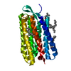
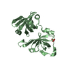
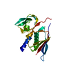
| Title | Structure of the C-terminal dimerization domain of infectious bronchitis virus nucleocapsid protein | ||||||
 Components Components | Nucleocapsid protein | ||||||
 Keywords Keywords | VIRUS/VIRAL PROTEIN/RNA BINDING PROTEIN / Nucleocapsid protein / N protein / Coronavirus / IBV N protein / Dimerization domain / VIRUS-VIRAL PROTEIN-RNA BINDING PROTEIN COMPLEX | ||||||
| Function / homology |  Function and homology information Function and homology informationviral nucleocapsid / host cell endoplasmic reticulum-Golgi intermediate compartment / host cell Golgi apparatus / ribonucleoprotein complex / DNA-templated transcription / RNA binding / identical protein binding Similarity search - Function | ||||||
| Biological species |  Infectious bronchitis virus Infectious bronchitis virus | ||||||
| Method |  X-RAY DIFFRACTION / SYNCHROTRON / MOLECULAR REPLACEMENT / Resolution: 2.2 Å X-RAY DIFFRACTION / SYNCHROTRON / MOLECULAR REPLACEMENT / Resolution: 2.2 Å | ||||||
 Authors Authors | Jayaram, H. / Fan, H. / Bowman, B.R. / Ooi, A. / Jayaram, J. / Collisson, E.W. / Lescar, J. / Prasad, B.V. | ||||||
 Citation Citation | Journal: J.Virol. / Year: 2006 Title: X-ray structures of the N- and C-terminal domains of a coronavirus nucleocapsid protein: implications for nucleocapsid formation. Authors: Jayaram, H. / Fan, H. / Bowman, B.R. / Ooi, A. / Jayaram, J. / Collisson, E.W. / Lescar, J. / Prasad, B.V. | ||||||
| History |
|
- Structure visualization
| Structure viewer | Molecule: MolmilJmol/JSmol |
|---|
- Downloads & links
-Download
| PDBx/mmCIF format | 2ge8.cif.gz | 172.5 KB | Display | PDBx/mmCIF format |
|---|---|---|---|---|
| PDB format | pdb2ge8.ent.gz | 141.7 KB | Display | PDB format |
| PDBx/mmJSON format | 2ge8.json.gz | Tree view | PDBx/mmJSON format | |
| Others |  Other downloads Other downloads |
-Validation report
| Arichive directory | https://data.pdbj.org/pub/pdb/validation_reports/ge/2ge8ftp://data.pdbj.org/pub/pdb/validation_reports/ge/2ge8 | HTTPS FTP |
|---|
-Related structure data
| Related structure data |  2c86C  2ca1C  2ge7SC  2gecC S: Starting model for refinement C: citing same article ( |
|---|---|
| Similar structure data |


-Links
 PDBj
PDBj- Assembly
| Deposited unit | 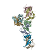
| ||||||||
|---|---|---|---|---|---|---|---|---|---|
| 1 | 
| ||||||||
| 2 | 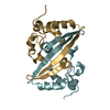
| ||||||||
| 3 | 
| ||||||||
| 4 | 
| ||||||||
| Unit cell |
| ||||||||
| Details | Dimer in solution and 4 dimers in assymetric unit. Hypothesized dimer-dimer interaction to form linaer arrays seen in assymetric unit with 8 molecules |
-Components
| #1: Protein | Mass: 12849.602 Da / Num. of mol.: 8 / Fragment: C-terminal domain Source method: isolated from a genetically manipulated source Source: (gene. exp.) Infectious bronchitis virus / Genus: Coronavirus / Strain: Gray / Gene: N / Plasmid: pET 41 Ek-LIC / Species (production host): Escherichia coli / Production host:  |
|---|
-Experimental details
-Experiment
| Experiment | Method: X-RAY DIFFRACTION / Number of used crystals: 1 |
|---|
- Sample preparation
| Crystal | Density Matthews: 2.43 Å3/Da / Density % sol: 49.42 % |
|---|---|
| Crystal grow | Temperature: 298 K / Method: vapor diffusion, hanging drop / pH: 8.6 Details: 30% PEG 4000, 100 mM Tris-HCl pH 8.6, 800 mM LiCl, VAPOR DIFFUSION, HANGING DROP, temperature 298K |
-Data collection
| Diffraction | Mean temperature: 178 K |
|---|---|
| Diffraction source | Source: SYNCHROTRON / Site: APS  / Beamline: 14-BM-C / Wavelength: 0.9 Å / Beamline: 14-BM-C / Wavelength: 0.9 Å |
| Detector | Type: ADSC QUANTUM 4 / Detector: CCD / Date: Dec 19, 2003 Details: Bent conical Si-mirror (Rh coated). Bent Ge(111) monochromator |
| Radiation | Monochromator: Bent Ge(111) monochromator / Protocol: SINGLE WAVELENGTH / Monochromatic (M) / Laue (L): M / Scattering type: x-ray |
| Radiation wavelength | Wavelength: 0.9 Å / Relative weight: 1 |
| Reflection | Resolution: 2→129.473 Å / Num. all: 69504 / Num. obs: 66258 / % possible obs: 95.8 % / Observed criterion σ(F): 2 / Observed criterion σ(I): 2 / Redundancy: 4.4 % / Rmerge(I) obs: 0.115 / Rsym value: 0.115 / Net I/σ(I): 4.8 |
| Reflection shell | Resolution: 2→2.11 Å / % possible obs: 97.3 % / Redundancy: 3.6 % / Rmerge(I) obs: 0.013 / Mean I/σ(I) obs: 0.5 / Num. measured all: 35383 / Num. unique obs: 9713 / Rsym value: 0.01276 |
- Processing
| Software |
| ||||||||||||||||||||||||||||||||||||||||||||||||||||||||||||||||||||||||||||||||||||||||||
|---|---|---|---|---|---|---|---|---|---|---|---|---|---|---|---|---|---|---|---|---|---|---|---|---|---|---|---|---|---|---|---|---|---|---|---|---|---|---|---|---|---|---|---|---|---|---|---|---|---|---|---|---|---|---|---|---|---|---|---|---|---|---|---|---|---|---|---|---|---|---|---|---|---|---|---|---|---|---|---|---|---|---|---|---|---|---|---|---|---|---|---|
| Refinement | Method to determine structure: MOLECULAR REPLACEMENT Starting model: PDB ENTRY 2GE7 4 copies Resolution: 2.2→47.78 Å / Cor.coef. Fo:Fc: 0.922 / Cor.coef. Fo:Fc free: 0.884 / SU B: 7.789 / SU ML: 0.197 / Cross valid method: THROUGHOUT / σ(F): 0 / σ(I): 2 / ESU R: 0.309 / ESU R Free: 0.247 / Stereochemistry target values: MAXIMUM LIKELIHOOD
| ||||||||||||||||||||||||||||||||||||||||||||||||||||||||||||||||||||||||||||||||||||||||||
| Solvent computation | Ion probe radii: 0.8 Å / Shrinkage radii: 0.8 Å / VDW probe radii: 1.2 Å / Solvent model: BABINET MODEL WITH MASK | ||||||||||||||||||||||||||||||||||||||||||||||||||||||||||||||||||||||||||||||||||||||||||
| Displacement parameters | Biso mean: 32.178 Å2
| ||||||||||||||||||||||||||||||||||||||||||||||||||||||||||||||||||||||||||||||||||||||||||
| Refinement step | Cycle: LAST / Resolution: 2.2→47.78 Å
| ||||||||||||||||||||||||||||||||||||||||||||||||||||||||||||||||||||||||||||||||||||||||||
| Refine LS restraints |
| ||||||||||||||||||||||||||||||||||||||||||||||||||||||||||||||||||||||||||||||||||||||||||
| LS refinement shell | Resolution: 2.199→2.256 Å / Total num. of bins used: 20
|
