 Movie
Movie Controller

[English] 日本語
 Yorodumi
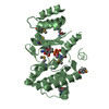
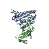
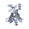
- PDB-2ge7: Structure of the C-terminal dimerization domain of infectious bro... -
+ Open data

- Basic information
| Entry | Database: PDB / ID: 2ge7 | ||||||
|---|---|---|---|---|---|---|---|
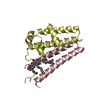
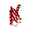
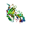
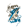
| Title | Structure of the C-terminal dimerization domain of infectious bronchitis virus nucleocapsid protein | ||||||
 Components Components | Nucleocapsid protein | ||||||
 Keywords Keywords | VIRUS/VIRAL PROTEIN/RNA BINDING PROTEIN / Nucleocapsid protein / N protein / Coronavirus / IBV N protein / Dimerization domain / VIRUS-VIRAL PROTEIN-RNA BINDING PROTEIN COMPLEX | ||||||
| Function / homology |  Function and homology information Function and homology informationviral nucleocapsid / host cell endoplasmic reticulum-Golgi intermediate compartment / host cell Golgi apparatus / ribonucleoprotein complex / DNA-templated transcription / RNA binding / identical protein binding Similarity search - Function | ||||||
| Biological species |  Infectious bronchitis virus Infectious bronchitis virus | ||||||
| Method |  X-RAY DIFFRACTION / SYNCHROTRON / MAD / Resolution: 2 Å X-RAY DIFFRACTION / SYNCHROTRON / MAD / Resolution: 2 Å | ||||||
 Authors Authors | Jayaram, H. / Fan, H. / Bowman, B.R. / Ooi, A. / Jayaram, J. / Collisson, E.W. / Lescar, J. / Prasad, B.V. | ||||||
 Citation Citation | Journal: J.Virol. / Year: 2006 Title: X-ray structures of the N- and C-terminal domains of a coronavirus nucleocapsid protein: implications for nucleocapsid formation. Authors: Jayaram, H. / Fan, H. / Bowman, B.R. / Ooi, A. / Jayaram, J. / Collisson, E.W. / Lescar, J. / Prasad, B.V. | ||||||
| History |
|
- Structure visualization
| Structure viewer | Molecule: MolmilJmol/JSmol |
|---|
- Downloads & links
-Download
| PDBx/mmCIF format | 2ge7.cif.gz | 54.6 KB | Display | PDBx/mmCIF format |
|---|---|---|---|---|
| PDB format | pdb2ge7.ent.gz | 40.9 KB | Display | PDB format |
| PDBx/mmJSON format | 2ge7.json.gz | Tree view | PDBx/mmJSON format | |
| Others |  Other downloads Other downloads |
-Validation report
| Arichive directory | https://data.pdbj.org/pub/pdb/validation_reports/ge/2ge7ftp://data.pdbj.org/pub/pdb/validation_reports/ge/2ge7 | HTTPS FTP |
|---|
-Related structure data


-Links
 PDBj
PDBj- Assembly
| Deposited unit | 
| ||||||||
|---|---|---|---|---|---|---|---|---|---|
| 1 |
| ||||||||
| Unit cell |
| ||||||||
| Details | Dimer in solution and in the assymetric unit |
-Components
| #1: Protein | Mass: 12107.784 Da / Num. of mol.: 2 / Fragment: C-terminal domain Source method: isolated from a genetically manipulated source Source: (gene. exp.) Infectious bronchitis virus / Genus: Coronavirus / Strain: Gray / Gene: N / Plasmid: pET 41 Ek-LIC / Species (production host): Escherichia coli / Production host:  #2: Water | ChemComp-HOH / |  Mass: 18.015 Da / Num. of mol.: 92 / Source method: isolated from a natural source / Formula: H2O Mass: 18.015 Da / Num. of mol.: 92 / Source method: isolated from a natural source / Formula: H2O |
|---|
-Experimental details
-Experiment
| Experiment | Method: X-RAY DIFFRACTION / Number of used crystals: 1 |
|---|
- Sample preparation
| Crystal | Density Matthews: 2.26 Å3/Da / Density % sol: 45.61 % |
|---|---|
| Crystal grow | Temperature: 298 K / Method: vapor diffusion, hanging drop / pH: 4.8 Details: PEG 4000, 28.75% to 29.5 %, pH 4.8 Citrate, 0.1 M MgCl2, VAPOR DIFFUSION, HANGING DROP, temperature 298.0K |
-Data collection
| Diffraction | Mean temperature: 178 K | |||||||||
|---|---|---|---|---|---|---|---|---|---|---|
| Diffraction source | Source: SYNCHROTRON / Site: APS  / Beamline: 19-ID / Wavelength: 0.97937 , 0.9795 / Beamline: 19-ID / Wavelength: 0.97937 , 0.9795 | |||||||||
| Detector | Type: SBC-2 / Detector: CCD / Date: Jun 24, 2004 Details: Rosenbaum-Rock monochromator #1 high-resolution double-crystal sagittal focusing, Rosenbaum-Rock monochromator #2 double crystal, Rosenbaum-Rock vertical focusing mirror | |||||||||
| Radiation | Monochromator: Rosenbaum-Rock monochromator / Protocol: MAD / Monochromatic (M) / Laue (L): M / Scattering type: x-ray | |||||||||
| Radiation wavelength |
| |||||||||
| Reflection | Resolution: 1.984→53.452 Å / Num. obs: 15780 / % possible obs: 96 % / Observed criterion σ(F): 2 / Observed criterion σ(I): 2 / Redundancy: 6.4 % / Limit h max: 18 / Limit h min: 0 / Limit k max: 33 / Limit k min: 0 / Limit l max: 45 / Limit l min: 0 / Rmerge(I) obs: 0.072 / Rsym value: 0.072 / Net I/σ(I): 28.8 | |||||||||
| Reflection shell | Resolution: 1.9→2 Å / Rmerge(I) obs: 0.282 / Mean I/σ(I) obs: 0.73 / Rsym value: 0.282 / % possible all: 95 |
- Processing
| Software |
| ||||||||||||||||||||||||||||||||||||||||||||||||||||||||||||||||||||||||||||||||||||||||||
|---|---|---|---|---|---|---|---|---|---|---|---|---|---|---|---|---|---|---|---|---|---|---|---|---|---|---|---|---|---|---|---|---|---|---|---|---|---|---|---|---|---|---|---|---|---|---|---|---|---|---|---|---|---|---|---|---|---|---|---|---|---|---|---|---|---|---|---|---|---|---|---|---|---|---|---|---|---|---|---|---|---|---|---|---|---|---|---|---|---|---|---|
| Refinement | Method to determine structure: MAD / Resolution: 2→53.45 Å / Cor.coef. Fo:Fc: 0.946 / Cor.coef. Fo:Fc free: 0.912 / SU B: 3.922 / SU ML: 0.112 / Cross valid method: THROUGHOUT / σ(F): 0 / σ(I): 2 / ESU R: 0.193 / ESU R Free: 0.183 / Stereochemistry target values: MAXIMUM LIKELIHOOD
| ||||||||||||||||||||||||||||||||||||||||||||||||||||||||||||||||||||||||||||||||||||||||||
| Solvent computation | Ion probe radii: 0.8 Å / Shrinkage radii: 0.8 Å / VDW probe radii: 1.2 Å / Solvent model: BABINET MODEL WITH MASK | ||||||||||||||||||||||||||||||||||||||||||||||||||||||||||||||||||||||||||||||||||||||||||
| Displacement parameters | Biso mean: 20.492 Å2
| ||||||||||||||||||||||||||||||||||||||||||||||||||||||||||||||||||||||||||||||||||||||||||
| Refinement step | Cycle: LAST / Resolution: 2→53.45 Å
| ||||||||||||||||||||||||||||||||||||||||||||||||||||||||||||||||||||||||||||||||||||||||||
| Refine LS restraints |
| ||||||||||||||||||||||||||||||||||||||||||||||||||||||||||||||||||||||||||||||||||||||||||
| LS refinement shell | Resolution: 2.005→2.057 Å / Total num. of bins used: 20
|
