[English] 日本語
 Yorodumi
- PDB-2gbh: NMR structure of stem region of helix-35 of 23S E.coli ribosomal ... -
+ Open data
- Basic information
| Entry | Database: PDB / ID: 2gbh | ||||||||||||||||||
|---|---|---|---|---|---|---|---|---|---|---|---|---|---|---|---|---|---|---|---|
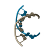
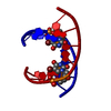
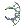
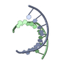
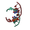
| Title | NMR structure of stem region of helix-35 of 23S E.coli ribosomal RNA (residues 736-760) | ||||||||||||||||||
 Components Components | 5'-R(* Keywords KeywordsRNA / RDC / rCSA | Function / homology | RNA / RNA (> 10) |  Function and homology information Function and homology informationMethod | SOLUTION NMR / Cartesian simulated annealing |  Authors AuthorsBax, A. / Boisbouvier, J. / Bryce, D. / Grishaev, A. / Jaroniec, C. / Miclet, E. / Nikonovicz, E. / O'Neil-Cabello, E. / Ying, J. |  Citation Citation Journal: J.Am.Chem.Soc. / Year: 2004 Journal: J.Am.Chem.Soc. / Year: 2004Title: Measurement of five dipolar couplings from a single 3D NMR multiplet applied to the study of RNA dynamics. Authors: O'Neil-Cabello, E. / Bryce, D.L. / Nikonowicz, E.P. / Bax, A. History |
|
- Structure visualization
| Structure viewer | Molecule:  Molmil Molmil Jmol/JSmol Jmol/JSmol |
|---|
- Downloads & links
- Download
| PDBx/mmCIF format |  2gbh.cif.gz 2gbh.cif.gz | 57.2 KB | Display |  PDBx/mmCIF format PDBx/mmCIF format |
|---|---|---|---|---|
| PDB format |  pdb2gbh.ent.gz pdb2gbh.ent.gz | 43.9 KB | Display |  PDB format PDB format |
| PDBx/mmJSON format |  2gbh.json.gz 2gbh.json.gz | Tree view |  PDBx/mmJSON format PDBx/mmJSON format | |
| Others |  Other downloads Other downloads |
-Validation report
| Arichive directory |  https://data.pdbj.org/pub/pdb/validation_reports/gb/2gbh https://data.pdbj.org/pub/pdb/validation_reports/gb/2gbh ftp://data.pdbj.org/pub/pdb/validation_reports/gb/2gbh ftp://data.pdbj.org/pub/pdb/validation_reports/gb/2gbh | HTTPS FTP |
|---|
-Related structure data
| Similar structure data |
|---|
- Links
Links
- Assembly
| Deposited unit | 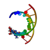
| |||||||||
|---|---|---|---|---|---|---|---|---|---|---|
| 1 |
| |||||||||
| NMR ensembles |
|
- Components
| #1: RNA chain | Mass: 7637.666 Da / Num. of mol.: 1 / Source method: obtained synthetically Details: The sequence of this RNA naturally exists in 23S Ribosomal RNA of E.Coli. |
|---|
-Experimental details
-Experiment
| Experiment | Method: SOLUTION NMR |
|---|---|
| NMR experiment | Type: RDC measurement |
- Sample preparation
| Details | Contents: 1.5mM helix-35psi U-15N,13C; 17mM NaCl, 17mM phosphate buffer; 99% D2O Solvent system: 99% D2O |
|---|---|
| Sample conditions | Ionic strength: 17 mM NaCl, 17 mM phosphate / pH: 6.8 / Pressure: ambient / Temperature: 298 K |
-NMR measurement
| Radiation | Protocol: SINGLE WAVELENGTH / Monochromatic (M) / Laue (L): M | |||||||||||||||
|---|---|---|---|---|---|---|---|---|---|---|---|---|---|---|---|---|
| Radiation wavelength | Relative weight: 1 | |||||||||||||||
| NMR spectrometer |
|
- Processing
| NMR software | Name:  Xplor-NIH / Version: 2.9.4 / Developer: Schwieters, Kuszewski, Tjandra, Clore / Classification: refinement Xplor-NIH / Version: 2.9.4 / Developer: Schwieters, Kuszewski, Tjandra, Clore / Classification: refinement |
|---|---|
| Refinement | Method: Cartesian simulated annealing / Software ordinal: 1 Details: Experimental restraints for the helix-35 stem are 277 RDCs, 13 31P anisotropic shifts, 41 dihedral angles and 188 NOEs. During the refinement the geometries of selected ribose rings were ...Details: Experimental restraints for the helix-35 stem are 277 RDCs, 13 31P anisotropic shifts, 41 dihedral angles and 188 NOEs. During the refinement the geometries of selected ribose rings were kept identical with the NCS restraint terms. Attractive non-bonded potentials were employed. No database-derived terms were used. The parameter file was modified with nucleotide type-specific values for distances, angles and improper torsions. Cross-validation statistics on the groups of 1/4 of ribose 1-bond C-H RDCs corresponds to an average Q-factor of 0.135. |
| NMR representative | Selection criteria: lowest energy |
| NMR ensemble | Conformer selection criteria: all calculated structures submitted Conformers calculated total number: 5 / Conformers submitted total number: 5 |
 Movie
Movie Controller




 PDBj
PDBj


