 Movie
Movie Controller

[English] 日本語
 Yorodumi
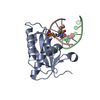
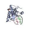
- PDB-2g8u: B. halodurans RNase H catalytic domain D132N mutant in complex wi... -
+ Open data

- Basic information
| Entry | Database: PDB / ID: 2g8u | ||||||
|---|---|---|---|---|---|---|---|
| Title | B. halodurans RNase H catalytic domain D132N mutant in complex with Mg2+ and RNA/DNA hybrid (non-P nick at the active site) | ||||||
 Components Components |
| ||||||
 Keywords Keywords | HYDROLASE/RNA/DNA / RNase H / Ribonuclease H / RNA/DNA hybrid / HYDROLASE-RNA-DNA COMPLEX | ||||||
| Function / homology |  Function and homology information Function and homology informationDNA replication, removal of RNA primer / ribonuclease H / RNA-DNA hybrid ribonuclease activity / nucleic acid binding / metal ion binding / cytoplasm Similarity search - Function | ||||||
| Biological species |  Bacillus halodurans (bacteria) Bacillus halodurans (bacteria) | ||||||
| Method |  X-RAY DIFFRACTION / MOLECULAR REPLACEMENT / Resolution: 2.7 Å X-RAY DIFFRACTION / MOLECULAR REPLACEMENT / Resolution: 2.7 Å | ||||||
 Authors Authors | Nowotny, M. / Yang, W. | ||||||
 Citation Citation | Journal: Embo J. / Year: 2006 Title: Stepwise analyses of metal ions in RNase H catalysis from substrate destabilization to product release. Authors: Nowotny, M. / Yang, W. | ||||||
| History |
|
- Structure visualization
| Structure viewer | Molecule: MolmilJmol/JSmol |
|---|
- Downloads & links
-Download
| PDBx/mmCIF format | 2g8u.cif.gz | 52.4 KB | Display | PDBx/mmCIF format |
|---|---|---|---|---|
| PDB format | pdb2g8u.ent.gz | 32.7 KB | Display | PDB format |
| PDBx/mmJSON format | 2g8u.json.gz | Tree view | PDBx/mmJSON format | |
| Others |  Other downloads Other downloads |
-Validation report
| Arichive directory | https://data.pdbj.org/pub/pdb/validation_reports/g8/2g8uftp://data.pdbj.org/pub/pdb/validation_reports/g8/2g8u | HTTPS FTP |
|---|
-Related structure data
| Related structure data |  2g8fC  2g8hC  2g8iC  2g8kC  2g8vC  2g8wC  1zbiS S: Starting model for refinement C: citing same article ( |
|---|---|
| Similar structure data |


-Links
 PDBj
PDBj
- Assembly
| Deposited unit | 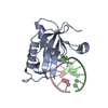
| ||||||||
|---|---|---|---|---|---|---|---|---|---|
| 1 |
| ||||||||
| Unit cell |
|
-Components
| #1: RNA chain | Mass: 1875.189 Da / Num. of mol.: 1 / Source method: obtained synthetically | ||
|---|---|---|---|
| #2: DNA chain | Mass: 1824.228 Da / Num. of mol.: 1 / Source method: obtained synthetically | ||
| #3: Protein | Mass: 16329.478 Da / Num. of mol.: 1 / Fragment: Bh-RNase HC / Mutation: D132N Source method: isolated from a genetically manipulated source Source: (gene. exp.) Bacillus halodurans (bacteria) / Gene: rnhA / Production host: | ||
| #4: Chemical |   Mass: 24.305 Da / Num. of mol.: 2 / Source method: obtained synthetically / Formula: Mg Mass: 24.305 Da / Num. of mol.: 2 / Source method: obtained synthetically / Formula: Mg#5: Water | ChemComp-HOH / |  Mass: 18.015 Da / Num. of mol.: 20 / Source method: isolated from a natural source / Formula: H2O Mass: 18.015 Da / Num. of mol.: 20 / Source method: isolated from a natural source / Formula: H2O |
-Experimental details
-Experiment
| Experiment | Method: X-RAY DIFFRACTION / Number of used crystals: 1 |
|---|
- Sample preparation
| Crystal | Density Matthews: 2.33 Å3/Da / Density % sol: 47.25 % | ||||||||||||||||||||||||||||||||
|---|---|---|---|---|---|---|---|---|---|---|---|---|---|---|---|---|---|---|---|---|---|---|---|---|---|---|---|---|---|---|---|---|---|
| Crystal grow | Temperature: 278 K / Method: vapor diffusion, sitting drop / pH: 7.5 Details: 25% MPD, 0.2M NaCl, 0.1M HEPES pH 7.5, VAPOR DIFFUSION, SITTING DROP, temperature 278K | ||||||||||||||||||||||||||||||||
| Components of the solutions |
|
-Data collection
| Diffraction | Mean temperature: 95 K |
|---|---|
| Diffraction source | Source: ROTATING ANODE / Type: RIGAKU |
| Detector | Type: RIGAKU RAXIS IV / Detector: IMAGE PLATE / Date: Jun 6, 2005 |
| Radiation | Protocol: SINGLE WAVELENGTH / Monochromatic (M) / Laue (L): M / Scattering type: x-ray |
| Radiation wavelength | Relative weight: 1 |
| Reflection | Resolution: 2.7→30 Å / Num. all: 5252 / Num. obs: 4718 / % possible obs: 89.8 % / Observed criterion σ(I): 2.4 |
| Reflection shell | Resolution: 2.7→2.82 Å |
- Processing
| Software |
| ||||||||||||||||||||
|---|---|---|---|---|---|---|---|---|---|---|---|---|---|---|---|---|---|---|---|---|---|
| Refinement | Method to determine structure: MOLECULAR REPLACEMENT Starting model: PDB ENTRY 1ZBI Resolution: 2.7→30 Å / σ(I): 2.4 / Stereochemistry target values: Engh & Huber
| ||||||||||||||||||||
| Refinement step | Cycle: LAST / Resolution: 2.7→30 Å
| ||||||||||||||||||||
| Refine LS restraints |
|
