 Movie
Movie Controller

[English] 日本語
 Yorodumi
- PDB-1md6: High resolution crystal structure of murine IL-1F5 reveals unique... -
+ Open data

- Basic information
| Entry | Database: PDB / ID: 1md6 | ||||||
|---|---|---|---|---|---|---|---|
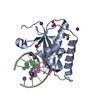
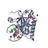
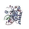
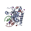
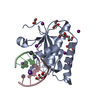
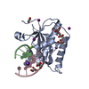
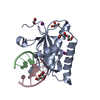
| Title | High resolution crystal structure of murine IL-1F5 reveals unique loop conformation for specificity | ||||||
 Components Components | interleukin 1 family, member 5 (delta) | ||||||
 Keywords Keywords | IMMUNE SYSTEM / beta triple / alpha helix | ||||||
| Function / homology |  Function and homology information Function and homology informationInterleukin-36 pathway / negative regulation of cytokine-mediated signaling pathway / antifungal humoral response / negative regulation of interleukin-17 production / interleukin-1 receptor binding / negative regulation of type II interferon production / negative regulation of interleukin-6 production / cytokine activity / cellular response to lipopolysaccharide / immune response ...Interleukin-36 pathway / negative regulation of cytokine-mediated signaling pathway / antifungal humoral response / negative regulation of interleukin-17 production / interleukin-1 receptor binding / negative regulation of type II interferon production / negative regulation of interleukin-6 production / cytokine activity / cellular response to lipopolysaccharide / immune response / inflammatory response / innate immune response / : / cytoplasm Similarity search - Function | ||||||
| Biological species |  | ||||||
| Method |  X-RAY DIFFRACTION / SYNCHROTRON / MOLECULAR REPLACEMENT / Resolution: 1.6 Å X-RAY DIFFRACTION / SYNCHROTRON / MOLECULAR REPLACEMENT / Resolution: 1.6 Å | ||||||
 Authors Authors | Pei, X.Y. / Dunn, E.F. / Gay, N.J. / O'Neill, L.A. | ||||||
 Citation Citation | Journal: Biochemistry / Year: 2003 Title: High-Resolution Structure of Murine Interleukin 1 Homologue IL-1F5 Reveals Unique Loop Conformations for Receptor Binding Specificity. Authors: Dunn, E.F. / Gay, N.J. / Bristow, A.F. / Gearing, D.P. / O'Neill, L.A. / Pei, X.Y. | ||||||
| History |
|
- Structure visualization
| Structure viewer | Molecule: MolmilJmol/JSmol |
|---|
- Downloads & links
-Download
| PDBx/mmCIF format | 1md6.cif.gz | 46.1 KB | Display | PDBx/mmCIF format |
|---|---|---|---|---|
| PDB format | pdb1md6.ent.gz | 32.1 KB | Display | PDB format |
| PDBx/mmJSON format | 1md6.json.gz | Tree view | PDBx/mmJSON format | |
| Others |  Other downloads Other downloads |
-Validation report
| Arichive directory | https://data.pdbj.org/pub/pdb/validation_reports/md/1md6ftp://data.pdbj.org/pub/pdb/validation_reports/md/1md6 | HTTPS FTP |
|---|
-Related structure data
| Related structure data |  2mibS S: Starting model for refinement |
|---|---|
| Similar structure data |


-Links
 PDBj
PDBj

- Assembly
| Deposited unit | 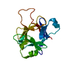
| ||||||||
|---|---|---|---|---|---|---|---|---|---|
| 1 |
| ||||||||
| Unit cell |
|
-Components
| #1: Protein | Mass: 16888.305 Da / Num. of mol.: 1 Source method: isolated from a genetically manipulated source Source: (gene. exp.)  |
|---|---|
| #2: Water | ChemComp-HOH /  Mass: 18.015 Da / Num. of mol.: 188 / Source method: isolated from a natural source / Formula: H2O Mass: 18.015 Da / Num. of mol.: 188 / Source method: isolated from a natural source / Formula: H2O |
| Has protein modification | Y |
-Experimental details
-Experiment
| Experiment | Method: X-RAY DIFFRACTION / Number of used crystals: 1 |
|---|
- Sample preparation
| Crystal | Density Matthews: 3.71 Å3/Da / Density % sol: 66.86 % | |||||||||||||||||||||||||||||||||||
|---|---|---|---|---|---|---|---|---|---|---|---|---|---|---|---|---|---|---|---|---|---|---|---|---|---|---|---|---|---|---|---|---|---|---|---|---|
| Crystal grow | Temperature: 291 K / Method: vapor diffusion, hanging drop / pH: 7.5 Details: ammomium sulphate, HEPES, potassium chloride, pH 7.5, VAPOR DIFFUSION, HANGING DROP, temperature 291K | |||||||||||||||||||||||||||||||||||
| Crystal grow | *PLUS Temperature: 18 ℃ / Method: vapor diffusion, hanging drop | |||||||||||||||||||||||||||||||||||
| Components of the solutions | *PLUS
|
-Data collection
| Diffraction | Mean temperature: 100 K |
|---|---|
| Diffraction source | Source: SYNCHROTRON / Site: ESRF  / Beamline: ID14-2 / Wavelength: 0.934 Å / Beamline: ID14-2 / Wavelength: 0.934 Å |
| Detector | Type: ADSC QUANTUM 4 / Detector: CCD / Date: Feb 28, 2002 / Details: mirrors |
| Radiation | Monochromator: Yale Mirrors / Protocol: SINGLE WAVELENGTH / Monochromatic (M) / Laue (L): M / Scattering type: x-ray |
| Radiation wavelength | Wavelength: 0.934 Å / Relative weight: 1 |
| Reflection | Resolution: 1.5→39.5 Å / Num. obs: 40586 / % possible obs: 100 % / Observed criterion σ(F): 1.4 / Observed criterion σ(I): 2.5 / Redundancy: 6.5 % / Biso Wilson estimate: 19.3 Å2 / Rmerge(I) obs: 0.085 / Rsym value: 0.078 / Net I/σ(I): 18.9 |
| Reflection shell | Resolution: 1.5→1.58 Å / Redundancy: 7 % / Rmerge(I) obs: 0.478 / Mean I/σ(I) obs: 3.7 / Num. unique all: 39005 / Rsym value: 0.478 / % possible all: 100 |
| Reflection | *PLUS Highest resolution: 1.58 Å / Num. obs: 40560 / Rmerge(I) obs: 0.078 |
| Reflection shell | *PLUS Redundancy: 7 % / Mean I/σ(I) obs: 1.4 |
- Processing
| Software |
| |||||||||||||||||||||||||
|---|---|---|---|---|---|---|---|---|---|---|---|---|---|---|---|---|---|---|---|---|---|---|---|---|---|---|
| Refinement | Method to determine structure: MOLECULAR REPLACEMENT Starting model: PDB ENTRY 2mib Resolution: 1.6→39.5 Å / Isotropic thermal model: anisotropic / Cross valid method: THROUGHOUT / σ(F): 0 / σ(I): 0 / Stereochemistry target values: Engh & Huber / Details: maximum likelihood target using amplitudes
| |||||||||||||||||||||||||
| Displacement parameters | Biso mean: 23 Å2
| |||||||||||||||||||||||||
| Refine analyze | Luzzati coordinate error obs: 0.13 Å / Luzzati d res low obs: 5 Å / Luzzati sigma a obs: 0.15 Å | |||||||||||||||||||||||||
| Refinement step | Cycle: LAST / Resolution: 1.6→39.5 Å
| |||||||||||||||||||||||||
| Refine LS restraints |
| |||||||||||||||||||||||||
| LS refinement shell | Resolution: 1.6→1.7 Å / Rfactor Rfree error: 0.016
| |||||||||||||||||||||||||
| Refinement | *PLUS Rfactor Rfree: 0.212 / Rfactor Rwork: 0.202 | |||||||||||||||||||||||||
| Solvent computation | *PLUS | |||||||||||||||||||||||||
| Displacement parameters | *PLUS | |||||||||||||||||||||||||
| Refine LS restraints | *PLUS
|
