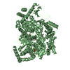
 Movie
Movie Controller

[English] 日本語
 Yorodumi
- PDB-2fzv: Crystal Structure of an apo form of a Flavin-binding Protein from... -
+ Open data

- Basic information
| Entry | Database: PDB / ID: 2fzv | ||||||
|---|---|---|---|---|---|---|---|
| Title | Crystal Structure of an apo form of a Flavin-binding Protein from Shigella flexneri | ||||||
 Components Components | putative arsenical resistance protein | ||||||
 Keywords Keywords | STRUCTURAL GENOMICS / UNKNOWN FUNCTION / Flavin binding Protein / PSI / Protein Structure Initiative / Midwest Center for Structural Genomics / MCSG | ||||||
| Function / homology |  Function and homology information Function and homology informationazobenzene reductase (NADP+) activity / FMN reductase (NADPH) activity / Oxidoreductases / oxidoreductase activity, acting on NAD(P)H, quinone or similar compound as acceptor / FMN binding / protein homotetramerization Similarity search - Function | ||||||
| Biological species |  Shigella flexneri 2a (bacteria) Shigella flexneri 2a (bacteria) | ||||||
| Method |  X-RAY DIFFRACTION / SYNCHROTRON / SIRAS / Resolution: 1.7 Å X-RAY DIFFRACTION / SYNCHROTRON / SIRAS / Resolution: 1.7 Å | ||||||
 Authors Authors | Vorontsov, I.I. / Minasov, G. / Brunzelle, J.S. / Shuvalova, L. / Collart, F.R. / Joachimiak, A. / Anderson, W.F. / Midwest Center for Structural Genomics (MCSG) | ||||||
 Citation Citation | Journal: Protein Sci. / Year: 2007 Title: Crystal structure of an apo form of Shigella flexneri ArsH protein with an NADPH-dependent FMN reductase activity Authors: Vorontsov, I.I. / Minasov, G. / Brunzelle, J.S. / Shuvalova, L. / Kiryukhina, O. / Collart, F.R. / Anderson, W.F. | ||||||
| History |
|
- Structure visualization
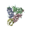
| Structure viewer | Molecule: MolmilJmol/JSmol |
|---|
- Downloads & links
-Download
| PDBx/mmCIF format | 2fzv.cif.gz | 231.6 KB | Display | PDBx/mmCIF format |
|---|---|---|---|---|
| PDB format | pdb2fzv.ent.gz | 185.2 KB | Display | PDB format |
| PDBx/mmJSON format | 2fzv.json.gz | Tree view | PDBx/mmJSON format | |
| Others |  Other downloads Other downloads |
-Validation report
| Arichive directory | https://data.pdbj.org/pub/pdb/validation_reports/fz/2fzvftp://data.pdbj.org/pub/pdb/validation_reports/fz/2fzv | HTTPS FTP |
|---|
-Related structure data
| Similar structure data | |
|---|---|
| Other databases |

-Links
 PDBj
PDBj- Assembly
| Deposited unit | 
| ||||||||
|---|---|---|---|---|---|---|---|---|---|
| 1 |
| ||||||||
| 2 | 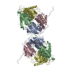
| ||||||||
| 3 | 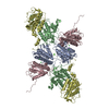
| ||||||||
| Unit cell |
| ||||||||
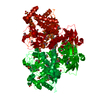
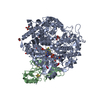
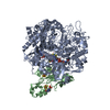
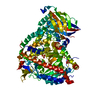
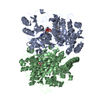
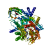
| Details | Chains A,B,C, and D represent biological assembly, which is homo-tetramer. |
-Components
| #1: Protein | Mass: 31179.279 Da / Num. of mol.: 4 Source method: isolated from a genetically manipulated source Source: (gene. exp.) Shigella flexneri 2a (bacteria) / Species: Shigella flexneri / Strain: 2457T / Gene: AAP17869 / Plasmid: pMCSG7 / Species (production host): Escherichia coli / Production host: #2: Chemical | ChemComp-CA / |   Mass: 40.078 Da / Num. of mol.: 1 / Source method: obtained synthetically / Formula: Ca Mass: 40.078 Da / Num. of mol.: 1 / Source method: obtained synthetically / Formula: Ca#3: Chemical |   Mass: 35.453 Da / Num. of mol.: 2 / Source method: obtained synthetically / Formula: Cl Mass: 35.453 Da / Num. of mol.: 2 / Source method: obtained synthetically / Formula: Cl#4: Water | ChemComp-HOH / |  Mass: 18.015 Da / Num. of mol.: 1111 / Source method: isolated from a natural source / Formula: H2O Mass: 18.015 Da / Num. of mol.: 1111 / Source method: isolated from a natural source / Formula: H2O |
|---|
-Experimental details
-Experiment
| Experiment | Method: X-RAY DIFFRACTION / Number of used crystals: 2 |
|---|
- Sample preparation
| Crystal | Density Matthews: 2.14 Å3/Da / Density % sol: 42.59 % |
|---|---|
| Crystal grow | Temperature: 295 K / Method: vapor diffusion, sitting drop / pH: 8.3 Details: 0.2 M Calcium chloride dihydrate, 20% w/v Polyethylene glycol 3350, 0.5 M NaCl, pH 8.3, VAPOR DIFFUSION, SITTING DROP, temperature 295K |
-Data collection
| Diffraction |
| |||||||||||||||
|---|---|---|---|---|---|---|---|---|---|---|---|---|---|---|---|---|
| Diffraction source |
| |||||||||||||||
| Detector |
| |||||||||||||||
| Radiation | Monochromator: Si 111 CHANNEL / Protocol: SINGLE WAVELENGTH / Monochromatic (M) / Laue (L): M / Scattering type: x-ray | |||||||||||||||
| Radiation wavelength |
| |||||||||||||||
| Reflection | Resolution: 1.7→29.81 Å / Num. all: 116180 / Num. obs: 116180 / % possible obs: 97.6 % / Observed criterion σ(I): -3 / Redundancy: 5.8 % / Rmerge(I) obs: 0.029 / Net I/σ(I): 38.33 | |||||||||||||||
| Reflection shell | Resolution: 1.7→1.8 Å / Redundancy: 3.6 % / Rmerge(I) obs: 0.24 / Mean I/σ(I) obs: 5.68 / Num. unique all: 17520 / % possible all: 94.7 |

- Processing
| Software |
| |||||||||||||||||||||||||||||||||||||||||||||||||||||||||||||||||||||||||||||||||||||||||||||||||||||||||||||||||||||||||||||
|---|---|---|---|---|---|---|---|---|---|---|---|---|---|---|---|---|---|---|---|---|---|---|---|---|---|---|---|---|---|---|---|---|---|---|---|---|---|---|---|---|---|---|---|---|---|---|---|---|---|---|---|---|---|---|---|---|---|---|---|---|---|---|---|---|---|---|---|---|---|---|---|---|---|---|---|---|---|---|---|---|---|---|---|---|---|---|---|---|---|---|---|---|---|---|---|---|---|---|---|---|---|---|---|---|---|---|---|---|---|---|---|---|---|---|---|---|---|---|---|---|---|---|---|---|---|---|
| Refinement | Method to determine structure: SIRAS / Resolution: 1.7→29.81 Å / Cor.coef. Fo:Fc: 0.958 / Cor.coef. Fo:Fc free: 0.931 / SU B: 6.595 / SU ML: 0.11 / TLS residual ADP flag: LIKELY RESIDUAL Isotropic thermal model: Atomic isotropic B-factor refinement Cross valid method: THROUGHOUT / σ(I): -3 / ESU R: 0.121 / ESU R Free: 0.123 / Stereochemistry target values: MAXIMUM LIKELIHOOD / Details: HYDROGENS HAVE BEEN ADDED IN THE RIDING POSITIONS
| |||||||||||||||||||||||||||||||||||||||||||||||||||||||||||||||||||||||||||||||||||||||||||||||||||||||||||||||||||||||||||||
| Solvent computation | Ion probe radii: 0.8 Å / Shrinkage radii: 0.8 Å / VDW probe radii: 1.2 Å / Solvent model: BABINET MODEL WITH MASK | |||||||||||||||||||||||||||||||||||||||||||||||||||||||||||||||||||||||||||||||||||||||||||||||||||||||||||||||||||||||||||||
| Displacement parameters | Biso mean: 23.033 Å2
| |||||||||||||||||||||||||||||||||||||||||||||||||||||||||||||||||||||||||||||||||||||||||||||||||||||||||||||||||||||||||||||
| Refinement step | Cycle: LAST / Resolution: 1.7→29.81 Å
| |||||||||||||||||||||||||||||||||||||||||||||||||||||||||||||||||||||||||||||||||||||||||||||||||||||||||||||||||||||||||||||
| Refine LS restraints |
| |||||||||||||||||||||||||||||||||||||||||||||||||||||||||||||||||||||||||||||||||||||||||||||||||||||||||||||||||||||||||||||
| LS refinement shell | Resolution: 1.7→1.745 Å / Total num. of bins used: 20
| |||||||||||||||||||||||||||||||||||||||||||||||||||||||||||||||||||||||||||||||||||||||||||||||||||||||||||||||||||||||||||||
| Refinement TLS params. | Method: refined / Refine-ID: X-RAY DIFFRACTION
| |||||||||||||||||||||||||||||||||||||||||||||||||||||||||||||||||||||||||||||||||||||||||||||||||||||||||||||||||||||||||||||
| Refinement TLS group |
|
