 Movie
Movie Controller


+ Open data

- Basic information
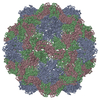
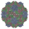
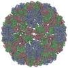
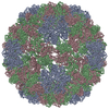
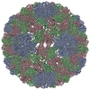
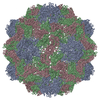
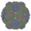
| Entry | Database: PDB / ID: 2fz2 | ||||||
|---|---|---|---|---|---|---|---|
| Title | Structure of Turnip Yellow Mosaic Virus at 100 K | ||||||
 Components Components |
| ||||||
 Keywords Keywords | Virus/RNA / Plant virus / Coat protein / Capsid protein / Tymoviruses / TYMV / RNA / Icosahedral virus / Virus-RNA COMPLEX | ||||||
| Function / homology |  Function and homology information Function and homology information | ||||||
| Biological species |  Turnip yellow mosaic virus Turnip yellow mosaic virus | ||||||
| Method |  X-RAY DIFFRACTION / SYNCHROTRON / MOLECULAR REPLACEMENT / Resolution: 2.9 Å X-RAY DIFFRACTION / SYNCHROTRON / MOLECULAR REPLACEMENT / Resolution: 2.9 Å | ||||||
 Authors Authors | Larson, S.B. / Lucas, R.W. / McPherson, A. | ||||||
 Citation Citation | Journal: Virology / Year: 2005 Title: The RNA of turnip yellow mosaic virus exhibits icosahedral order. Authors: Larson, S.B. / Lucas, R.W. / Greenwood, A. / McPherson, A. #1: Journal: Nat.Struct.Biol. / Year: 1996Title: Crystal structure of turnip yellow mosaic virus. Authors: Canady, M.A. / Larson, S.B. / Day, J. / McPherson, A. #2: Journal: Proteins / Year: 1995 Title: Preliminary X-ray diffraction analysis of crystals of turnip yellow mosaic virus (TYMV). Authors: Canady, M.A. / Day, J. / McPherson, A. #3: Journal: J.Mol.Biol. / Year: 2004Title: Crystal structure of an empty capsid of turnip yellow mosaic virus. Authors: van Roon, A.M. / Bink, H.H. / Plaisier, J.R. / Pleij, C.W. / Abrahams, J.P. / Pannu, N.S. | ||||||
| History |
|
- Structure visualization
| Structure viewer | Molecule: MolmilJmol/JSmol |
|---|
- Downloads & links
-Download
| PDBx/mmCIF format | 2fz2.cif.gz | 114.6 KB | Display | PDBx/mmCIF format |
|---|---|---|---|---|
| PDB format | pdb2fz2.ent.gz | 90.1 KB | Display | PDB format |
| PDBx/mmJSON format | 2fz2.json.gz | Tree view | PDBx/mmJSON format | |
| Others |  Other downloads Other downloads |
-Validation report
| Arichive directory | https://data.pdbj.org/pub/pdb/validation_reports/fz/2fz2ftp://data.pdbj.org/pub/pdb/validation_reports/fz/2fz2 | HTTPS FTP |
|---|
-Related structure data
| Related structure data |  2fz1C  1auyS S: Starting model for refinement C: citing same article ( |
|---|---|
| Similar structure data |


-Links
 PDBj
PDBj
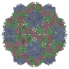
- Assembly
| Deposited unit | 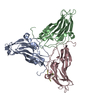
| ||||||||||||||||||||||||||||||||||||||||||||||||||||||||||||||||
|---|---|---|---|---|---|---|---|---|---|---|---|---|---|---|---|---|---|---|---|---|---|---|---|---|---|---|---|---|---|---|---|---|---|---|---|---|---|---|---|---|---|---|---|---|---|---|---|---|---|---|---|---|---|---|---|---|---|---|---|---|---|---|---|---|---|
| 1 | x 60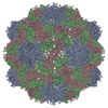
| ||||||||||||||||||||||||||||||||||||||||||||||||||||||||||||||||
| 2 |
| ||||||||||||||||||||||||||||||||||||||||||||||||||||||||||||||||
| 3 | x 5
| ||||||||||||||||||||||||||||||||||||||||||||||||||||||||||||||||
| 4 | x 6
| ||||||||||||||||||||||||||||||||||||||||||||||||||||||||||||||||
| 5 | 
| ||||||||||||||||||||||||||||||||||||||||||||||||||||||||||||||||
| 6 | x 15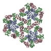
| ||||||||||||||||||||||||||||||||||||||||||||||||||||||||||||||||
| Unit cell |
| ||||||||||||||||||||||||||||||||||||||||||||||||||||||||||||||||
| Symmetry | Point symmetry: (Hermann–Mauguin notation: 532 / Schoenflies symbol: I (icosahedral)) | ||||||||||||||||||||||||||||||||||||||||||||||||||||||||||||||||
| Noncrystallographic symmetry (NCS) | NCS oper:
|
-Components
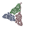
| #1: RNA chain | Mass: 870.588 Da / Num. of mol.: 1 / Source method: obtained synthetically |
|---|---|
| #2: Protein | Mass: 20207.285 Da / Num. of mol.: 3 / Fragment: Viral coat protein Source method: isolated from a genetically manipulated source Source: (gene. exp.) Turnip yellow mosaic virus / Genus: Tymovirus / Strain: (Australian isolate) / Species (production host): Brassica rapa / Production host:  |
-Experimental details
-Experiment
| Experiment | Method: X-RAY DIFFRACTION / Number of used crystals: 2 |
|---|
- Sample preparation
| Crystal | Density Matthews: 4.15 Å3/Da / Density % sol: 70.4 % |
|---|---|
| Crystal grow | Temperature: 298 K / Method: vapor diffusion, hanging drop / pH: 4.4 Details: 1.0-1.7 M NH4PO4, 0.1 M MES buffer, pH 4.4, VAPOR DIFFUSION, HANGING DROP, temperature 298K |
-Data collection
| Diffraction |
| ||||||||||||||||||
|---|---|---|---|---|---|---|---|---|---|---|---|---|---|---|---|---|---|---|---|
| Diffraction source |
| ||||||||||||||||||
| Detector |
| ||||||||||||||||||
| Radiation |
| ||||||||||||||||||
| Radiation wavelength |
| ||||||||||||||||||
| Reflection | Resolution: 2.61→59.12 Å / Num. all: 247650 / Num. obs: 225845 / % possible obs: 35.3 % / Observed criterion σ(F): 0 / Observed criterion σ(I): 0 / Redundancy: 3.2 % / Biso Wilson estimate: 34 Å2 / Rsym value: 0.106 / Net I/σ(I): 7.49 | ||||||||||||||||||
| Reflection shell | Resolution: 2.61→2.65 Å / Redundancy: 1 % / Mean I/σ(I) obs: -0.41 / Num. unique all: 172 / % possible all: 0.5 |

- Processing
| Software |
| ||||||||||||||||||||||||||||||||||||
|---|---|---|---|---|---|---|---|---|---|---|---|---|---|---|---|---|---|---|---|---|---|---|---|---|---|---|---|---|---|---|---|---|---|---|---|---|---|
| Refinement | Method to determine structure: MOLECULAR REPLACEMENT Starting model: PDB ENTRY 1AUY Resolution: 2.9→59.12 Å / Rfactor Rfree error: 0.003 / Data cutoff high absF: 171751.53 / Data cutoff low absF: 0 / Isotropic thermal model: RESTRAINED / Cross valid method: THROUGHOUT / σ(F): 2 / Stereochemistry target values: Engh & Huber Details: Simulated annealing and conjugate gradient minimization. Stereochemistry target values for the nucleic acid fragment are from Parkinson, Vojtechovsky, Clowney, Brunger & Berman
| ||||||||||||||||||||||||||||||||||||
| Solvent computation | Solvent model: FLAT MODEL / Bsol: 60.3842 Å2 / ksol: 0.394524 e/Å3 | ||||||||||||||||||||||||||||||||||||
| Displacement parameters | Biso mean: 29.3 Å2
| ||||||||||||||||||||||||||||||||||||
| Refine analyze |
| ||||||||||||||||||||||||||||||||||||
| Refinement step | Cycle: LAST / Resolution: 2.9→59.12 Å
| ||||||||||||||||||||||||||||||||||||
| Refine LS restraints |
| ||||||||||||||||||||||||||||||||||||
| LS refinement shell | Resolution: 2.9→3.08 Å / Rfactor Rfree error: 0.046 / Total num. of bins used: 6
| ||||||||||||||||||||||||||||||||||||
| Xplor file |
|
