Resolution: 3.4→50 Å / Rfactor Rfree error: 0.003 / Isotropic thermal model: RESTRAINED / Cross valid method: THROUGHOUT / σ(F): 2 / Stereochemistry target values: Engh & Huber Details: NCS RESTRAINTS WERE USED FOR BETA STRANDS AND HELICES OF EQUIVALENT SUBUNITS. IONS WERE FIXED AS RNA PHOSPHATES. ICOSAHEDRAL NCS CONSTRAINTS WERE APPLIED THROUGHOUT REFINEMENT.
Rfactor
Num. reflection
% reflection
Selection details
Rfree
0.228
6391
5 %
RANDOM
Rwork
0.218
-
-
-
all
0.278
156468
-
-
obs
-
127436
50.8 %
-
Displacement parameters
Biso mean: 105 Å2
Refine analyze
Free
Obs
Luzzati coordinate error
0.62 Å
0.55 Å
Luzzati d res low
-
5 Å
Luzzati sigma a
1.69 Å
1.61 Å
Refinement step
Cycle: LAST / Resolution: 3.4→50 Å
Protein
Nucleic acid
Ligand
Solvent
Total
Num. atoms
4239
63
6
0
4308
Refine LS restraints
Refine-ID
Type
Dev ideal
Dev ideal target
X-RAY DIFFRACTION
x_bond_d
0.009
X-RAY DIFFRACTION
x_bond_d_na
X-RAY DIFFRACTION
x_bond_d_prot
X-RAY DIFFRACTION
x_angle_d
X-RAY DIFFRACTION
x_angle_d_na
X-RAY DIFFRACTION
x_angle_d_prot
X-RAY DIFFRACTION
x_angle_deg
1.66
X-RAY DIFFRACTION
x_angle_deg_na
X-RAY DIFFRACTION
x_angle_deg_prot
X-RAY DIFFRACTION
x_dihedral_angle_d
29.3
X-RAY DIFFRACTION
x_dihedral_angle_d_na
X-RAY DIFFRACTION
x_dihedral_angle_d_prot
X-RAY DIFFRACTION
x_improper_angle_d
1.03
X-RAY DIFFRACTION
x_improper_angle_d_na
X-RAY DIFFRACTION
x_improper_angle_d_prot
X-RAY DIFFRACTION
x_mcbond_it
7.5
1.5
X-RAY DIFFRACTION
x_mcangle_it
12.25
2
X-RAY DIFFRACTION
x_scbond_it
16.25
2
X-RAY DIFFRACTION
x_scangle_it
22.85
2.5
Refine LS restraints NCS
NCS model details: CONSTRAINTS / Rms dev position: 0.059 Å / Weight Biso: 4 / Weight position: 100
LS refinement shell
Resolution: 3.4→3.52 Å / Rfactor Rfree error: 0.088 / Total num. of bins used: 10
Rfactor
Num. reflection
% reflection
Rfree
0.488
30
4.942 %
Rwork
0.402
607
-
obs
-
-
2.4 %
Xplor file
Refine-ID
Serial no
Param file
Topol file
X-RAY DIFFRACTION
1
PROTEIN_REP.PARAM
PROTEIN.TOP
X-RAY DIFFRACTION
2
DNA-RNA.PARAM
DNA-RNA.TOP
X-RAY DIFFRACTION
3
PO4_XPLOR_PAR.TXT
PO4_XPLOR_TOP.TXT
X-RAY DIFFRACTION
4
MG_XPLOR_PAR.TXT
MG_XPLOR_TOP.TXT
Refinement
*PLUS
Lowest resolution: 50 Å / % reflection Rfree: 5 %
Solvent computation
*PLUS
Displacement parameters
*PLUS
Refine LS restraints
*PLUS
Refine-ID
Type
Dev ideal
X-RAY DIFFRACTION
x_dihedral_angle_d
X-RAY DIFFRACTION
x_dihedral_angle_deg
29.3
X-RAY DIFFRACTION
x_improper_angle_d
X-RAY DIFFRACTION
x_improper_angle_deg
1.03
+
About Yorodumi
-
News
-
Feb 9, 2022. New format data for meta-information of EMDB entries
New format data for meta-information of EMDB entries
Version 3 of the EMDB header file is now the official format.
The previous official version 1.9 will be removed from the archive.
In the structure databanks used in Yorodumi, some data are registered as the other names, "COVID-19 virus" and "2019-nCoV". Here are the details of the virus and the list of structure data.
Jan 31, 2019. EMDB accession codes are about to change! (news from PDBe EMDB page)
EMDB accession codes are about to change! (news from PDBe EMDB page)
The allocation of 4 digits for EMDB accession codes will soon come to an end. Whilst these codes will remain in use, new EMDB accession codes will include an additional digit and will expand incrementally as the available range of codes is exhausted. The current 4-digit format prefixed with “EMD-” (i.e. EMD-XXXX) will advance to a 5-digit format (i.e. EMD-XXXXX), and so on. It is currently estimated that the 4-digit codes will be depleted around Spring 2019, at which point the 5-digit format will come into force.
The EM Navigator/Yorodumi systems omit the EMD- prefix.
Related info.:Q: What is EMD? / ID/Accession-code notation in Yorodumi/EM Navigator
Yorodumi is a browser for structure data from EMDB, PDB, SASBDB, etc.
This page is also the successor to EM Navigator detail page, and also detail information page/front-end page for Omokage search.
The word "yorodu" (or yorozu) is an old Japanese word meaning "ten thousand". "mi" (miru) is to see.
Related info.:EMDB / PDB / SASBDB / Comparison of 3 databanks / Yorodumi Search / Aug 31, 2016. New EM Navigator & Yorodumi / Yorodumi Papers / Jmol/JSmol / Function and homology information / Changes in new EM Navigator and Yorodumi
 Movie
Movie Controller


 Open data
 Basic information
 Components
 Keywords
 Function and homology information
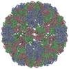
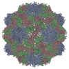
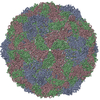
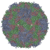
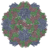
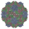
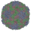
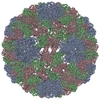
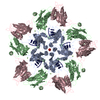
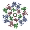
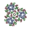
 Tomato aspermy virus
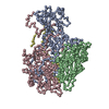
 X-RAY DIFFRACTION /
  Authors
 Citation
 Structure visualization
 Downloads & links
 Other downloads


 PDBj
PDBj
 Assembly
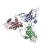

 Mass: 94.971 Da / Num. of mol.: 1 / Source method: obtained synthetically / Formula: PO4
 Mass: 24.305 Da / Num. of mol.: 1 / Source method: obtained synthetically / Formula: Mg
 Sample preparation
 Processing