[English] 日本語
Yorodumi
- PDB-1w39: Crystal structure of an artificial top component of turnip yellow... -

+
Open data


ID or keywords:

Loading...

-
Basic information

Entry
Database: PDB / ID: 1w39
TitleCrystal structure of an artificial top component of turnip yellow mosaic virus
ComponentsTURNIP YELLOW MOSAIC VIRUS EMPTY CAPSID
KeywordsVIRUS / ARTIFICIAL TOP COMPONENT / CAPSID PROTEIN / TYMV / VIRUS/VIRAL PROTEIN / ICOSAHEDRAL VIRUS
Function / homology
Function and homology information


T=3 icosahedral viral capsid / structural molecule activity
Similarity search - Function
Tymovirus coat protein / Tymovirus coat protein / Jelly Rolls - #20 / Viral coat protein subunit / Jelly Rolls / Sandwich / Mainly Beta
Similarity search - Domain/homology
Biological speciesTURNIP YELLOW MOSAIC VIRUS
MethodX-RAY DIFFRACTION / SYNCHROTRON / MOLECULAR REPLACEMENT / Resolution: 3.75 Å
Authorsvan Roon, A.M.M. / Bink, H.H.J. / Plaisier, J.R. / Pleij, C.W.A. / Abrahams, J.P. / Pannu, N.S.
Citation
Journal: J.Mol.Biol. / Year: 2004
Title: Crystal Structure of an Empty Capsid of Turnip Yellow Mosaic Virus.
Authors: Van Roon, A.M.M. / Bink, H.H.J. / Plaisier, J.R. / Pleij, C.W.A. / Abrahams, J.P. / Pannu, N.S.
#1: Journal: Nat.Struct.Biol. / Year: 1996
Title: Crystal Structure of Turnip Yellow Mosaic Virus
Authors: Canady, M.A. / Larson, S.B. / Day, J. / McPherson, A.
History
DepositionJul 14, 2004Deposition site: PDBE / Processing site: PDBE
Revision 1.0Aug 16, 2004Provider: repository / Type: Initial release
Revision 1.1May 8, 2011Group: Version format compliance
Revision 1.2Jul 13, 2011Group: Version format compliance
Revision 1.3Aug 9, 2017Group: Data collection / Category: diffrn_detector / Item: _diffrn_detector.type
Revision 1.4May 8, 2019Group: Data collection / Experimental preparation / Category: exptl_crystal_grow / Item: _exptl_crystal_grow.method
Revision 1.5Dec 13, 2023Group: Data collection / Database references ...Data collection / Database references / Derived calculations / Other / Refinement description
Category: chem_comp_atom / chem_comp_bond ...chem_comp_atom / chem_comp_bond / database_2 / pdbx_database_status / pdbx_initial_refinement_model / pdbx_struct_oper_list
Item: _database_2.pdbx_DOI / _database_2.pdbx_database_accession ..._database_2.pdbx_DOI / _database_2.pdbx_database_accession / _pdbx_database_status.status_code_sf / _pdbx_struct_oper_list.name / _pdbx_struct_oper_list.symmetry_operation / _pdbx_struct_oper_list.type
Remark 700 SHEET THE SHEET STRUCTURE OF THIS MOLECULE IS BIFURCATED. IN ORDER TO REPRESENT THIS FEATURE IN ... SHEET THE SHEET STRUCTURE OF THIS MOLECULE IS BIFURCATED. IN ORDER TO REPRESENT THIS FEATURE IN THE SHEET RECORDS BELOW, TWO SHEETS ARE DEFINED.

-
Structure visualization

Structure viewerMolecule:
MolmilJmol/JSmol

Downloads & links

-
Assembly

Deposited unit
A: TURNIP YELLOW MOSAIC VIRUS EMPTY CAPSID
B: TURNIP YELLOW MOSAIC VIRUS EMPTY CAPSID
C: TURNIP YELLOW MOSAIC VIRUS EMPTY CAPSID


Theoretical massNumber of molelcules
Total (without water)60,4303
Polymers60,4303
Non-polymers00
Water00
1
A: TURNIP YELLOW MOSAIC VIRUS EMPTY CAPSID
B: TURNIP YELLOW MOSAIC VIRUS EMPTY CAPSID
C: TURNIP YELLOW MOSAIC VIRUS EMPTY CAPSID
x 60


Theoretical massNumber of molelcules
Total (without water)3,625,784180
Polymers3,625,784180
Non-polymers00
Water0
TypeNameSymmetry operationNumber
identity operation1_555x,y,z1
point symmetry operation59
2


  • Idetical with deposited unit
  • icosahedral asymmetric unit
TypeNameSymmetry operationNumber
identity operation1_555x,y,z1
3
A: TURNIP YELLOW MOSAIC VIRUS EMPTY CAPSID
B: TURNIP YELLOW MOSAIC VIRUS EMPTY CAPSID
C: TURNIP YELLOW MOSAIC VIRUS EMPTY CAPSID
x 5


  • icosahedral pentamer
  • 302 kDa, 15 polymers
Theoretical massNumber of molelcules
Total (without water)302,14915
Polymers302,14915
Non-polymers00
Water0
TypeNameSymmetry operationNumber
identity operation1_555x,y,z1
point symmetry operation4
4
A: TURNIP YELLOW MOSAIC VIRUS EMPTY CAPSID
B: TURNIP YELLOW MOSAIC VIRUS EMPTY CAPSID
C: TURNIP YELLOW MOSAIC VIRUS EMPTY CAPSID
x 6


  • icosahedral 23 hexamer
  • 363 kDa, 18 polymers
Theoretical massNumber of molelcules
Total (without water)362,57818
Polymers362,57818
Non-polymers00
Water0
TypeNameSymmetry operationNumber
identity operation1_555x,y,z1
point symmetry operation5
5


  • Idetical with deposited unit in distinct coordinate
  • icosahedral asymmetric unit, std point frame
TypeNameSymmetry operationNumber
transform to point frame1
6
A: TURNIP YELLOW MOSAIC VIRUS EMPTY CAPSID
B: TURNIP YELLOW MOSAIC VIRUS EMPTY CAPSID
C: TURNIP YELLOW MOSAIC VIRUS EMPTY CAPSID
x 5


  • crystal asymmetric unit, crystal frame
  • 302 kDa, 15 polymers
Theoretical massNumber of molelcules
Total (without water)302,14915
Polymers302,14915
Non-polymers00
Water0
TypeNameSymmetry operationNumber
identity operation1_555x,y,z2
point symmetry operation4
Unit cell
Length a, b, c (Å)688.457, 688.457, 688.457
Angle α, β, γ (deg.)90.00, 90.00, 90.00
Int Tables number210
Space group name H-MF4132
SymmetryPoint symmetry: (Schoenflies symbol: I (icosahedral))
Noncrystallographic symmetry (NCS)NCS oper:
IDCodeMatrixVector
1given(1), (1), (1)
2generate(0.5, 0.309017, 0.809017), (0.309017, 0.809017, -0.5), (-0.809017, 0.5, 0.309017)-212.7449, 131.48358, -5.0E-5
3generate(-0.309017, 0.809017, 0.5), (0.809017, 0.5, -0.309017), (-0.5, 0.309017, -0.809017)-278.48673, 172.11427, 237.85597
4generate(-0.309017, 0.809017, -0.5), (0.809017, 0.5, 0.309017), (0.5, -0.309017, -0.809017)-106.37252, 65.74183, 384.85912
5generate(0.5, 0.309017, -0.809017), (0.309017, 0.809017, 0.5), (0.809017, -0.5, 0.309017)65.74175, -40.63064, 237.85605

-
Components

#1: Protein TURNIP YELLOW MOSAIC VIRUS EMPTY CAPSID


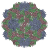
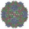
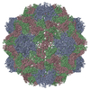
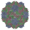
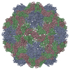
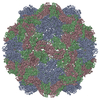
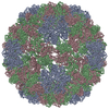
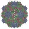
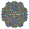
Mass: 20143.244 Da / Num. of mol.: 3 / Source method: isolated from a natural source
Details: COAT PROTEIN WAS REPORTED TO BE ACETYLATED AT THE N-TERMINUS OF CHAINS A, B, AND C, BUT NO N-TERMINAL ACETYL GROUP WAS OBSERVED
Source: (natural) TURNIP YELLOW MOSAIC VIRUS / References: UniProt: P03608
Sequence detailsRESIDUES 1-9 AND 16-26 OF CHAIN A WERE NOT OBSERVED IN THE ELECTRON DENSITY MAPS AND THEREFORE NOT MODELED

-
Experimental details

-
Experiment

ExperimentMethod: X-RAY DIFFRACTION / Number of used crystals: 1

-
Sample preparation

CrystalDensity Matthews: 11.3 Å3/Da / Density % sol: 89.1 % / Description: NONE
Crystal growMethod: vapor diffusion, sitting drop / pH: 4.5 / Details: 1.16 M AMMONIUM PHOSPHATE PH 4.5, SITTING DROP

-
Data collection

DiffractionMean temperature: 100 K
Diffraction sourceSource: SYNCHROTRON / Site: ESRF / Beamline: ID29 / Wavelength: 0.98
DetectorType: ADSC QUANTUM 210 / Detector: CCD / Date: Feb 15, 2003 / Details: MIRRORS
RadiationMonochromator: SI(311) MONOCHROMATOR / Protocol: SINGLE WAVELENGTH / Monochromatic (M) / Laue (L): M / Scattering type: x-ray
Radiation wavelengthWavelength: 0.98 Å / Relative weight: 1
ReflectionResolution: 3.75→25 Å / Num. obs: 140727 / % possible obs: 100 % / Observed criterion σ(I): 2 / Redundancy: 9 % / Biso Wilson estimate: 36.3 Å2 / Rmerge(I) obs: 0.2 / Net I/σ(I): 10.7
Reflection shellResolution: 3.75→3.88 Å / Redundancy: 6.3 % / Rmerge(I) obs: 0.6 / Mean I/σ(I) obs: 2.7 / % possible all: 99.3

-
Processing

Software
NameVersionClassification
CNS0.9refinement
DENZOdata reduction
SCALEPACKdata scaling
MOLREPphasing
RefinementMethod to determine structure: MOLECULAR REPLACEMENT
Starting model: PDB ENTRY 1AUY
Resolution: 3.75→25 Å / Rfactor Rfree error: 0.003 / Data cutoff high absF: 10000 / Isotropic thermal model: GROUP / Cross valid method: THROUGHOUT / σ(F): 0
RfactorNum. reflection% reflectionSelection details
Rfree0.2508 7052 5 %RANDOM
Rwork0.2453 ---
obs0.2453 140727 100 %-
Displacement parametersBiso mean: 50.6 Å2
Refine analyze
FreeObs
Luzzati coordinate error0.36 Å0.35 Å
Luzzati d res low-5 Å
Luzzati sigma a0.78 Å0.78 Å
Refinement stepCycle: LAST / Resolution: 3.75→25 Å
ProteinNucleic acidLigandSolventTotal
Num. atoms4094 0 0 0 4094
Refine LS restraints
Refine-IDTypeDev ideal
X-RAY DIFFRACTIONc_bond_d0.008294
X-RAY DIFFRACTIONc_bond_d_na
X-RAY DIFFRACTIONc_bond_d_prot
X-RAY DIFFRACTIONc_angle_d
X-RAY DIFFRACTIONc_angle_d_na
X-RAY DIFFRACTIONc_angle_d_prot
X-RAY DIFFRACTIONc_angle_deg1.48397
X-RAY DIFFRACTIONc_angle_deg_na
X-RAY DIFFRACTIONc_angle_deg_prot
X-RAY DIFFRACTIONc_dihedral_angle_d
X-RAY DIFFRACTIONc_dihedral_angle_d_na
X-RAY DIFFRACTIONc_dihedral_angle_d_prot
X-RAY DIFFRACTIONc_improper_angle_d
X-RAY DIFFRACTIONc_improper_angle_d_na
X-RAY DIFFRACTIONc_improper_angle_d_prot
X-RAY DIFFRACTIONc_mcbond_it
X-RAY DIFFRACTIONc_mcangle_it
X-RAY DIFFRACTIONc_scbond_it
X-RAY DIFFRACTIONc_scangle_it
Refine LS restraints NCSNCS model details: STRICT
LS refinement shellResolution: 3.75→3.98 Å / Rfactor Rfree error: 0.008 / Total num. of bins used: 8
RfactorNum. reflection% reflection
Rfree0.284 1183 5.1 %
Rwork0.286 21924 -
obs--99.8 %
Xplor fileSerial no: 1 / Param file: PROTEIN_REP.PARAM / Topol file: PROTEIN.TOP

+
About Yorodumi

-
News

-
Feb 9, 2022. New format data for meta-information of EMDB entries

New format data for meta-information of EMDB entries

  • Version 3 of the EMDB header file is now the official format.
  • The previous official version 1.9 will be removed from the archive.

Related info.:EMDB header

External links:wwPDB to switch to version 3 of the EMDB data model

-
Aug 12, 2020. Covid-19 info

Covid-19 info

URL: https://pdbjlvh1.pdbj.org/emnavi/covid19.php

New page: Covid-19 featured information page in EM Navigator.

Related info.:Covid-19 info / Mar 5, 2020. Novel coronavirus structure data

+
Mar 5, 2020. Novel coronavirus structure data

Novel coronavirus structure data

Related info.:Yorodumi Speices / Aug 12, 2020. Covid-19 info

External links:COVID-19 featured content - PDBj / Molecule of the Month (242):Coronavirus Proteases

+
Jan 31, 2019. EMDB accession codes are about to change! (news from PDBe EMDB page)

EMDB accession codes are about to change! (news from PDBe EMDB page)

  • The allocation of 4 digits for EMDB accession codes will soon come to an end. Whilst these codes will remain in use, new EMDB accession codes will include an additional digit and will expand incrementally as the available range of codes is exhausted. The current 4-digit format prefixed with “EMD-” (i.e. EMD-XXXX) will advance to a 5-digit format (i.e. EMD-XXXXX), and so on. It is currently estimated that the 4-digit codes will be depleted around Spring 2019, at which point the 5-digit format will come into force.
  • The EM Navigator/Yorodumi systems omit the EMD- prefix.

Related info.:Q: What is EMD? / ID/Accession-code notation in Yorodumi/EM Navigator

External links:EMDB Accession Codes are Changing Soon! / Contact to PDBj

+
Jul 12, 2017. Major update of PDB

Major update of PDB

  • wwPDB released updated PDB data conforming to the new PDBx/mmCIF dictionary.
  • This is a major update changing the version number from 4 to 5, and with Remediation, in which all the entries are updated.
  • In this update, many items about electron microscopy experimental information are reorganized (e.g. em_software).
  • Now, EM Navigator and Yorodumi are based on the updated data.

External links:wwPDB Remediation / Enriched Model Files Conforming to OneDep Data Standards Now Available in the PDB FTP Archive

-
Yorodumi

Thousand views of thousand structures

  • Yorodumi is a browser for structure data from EMDB, PDB, SASBDB, etc.
  • This page is also the successor to EM Navigator detail page, and also detail information page/front-end page for Omokage search.
  • The word "yorodu" (or yorozu) is an old Japanese word meaning "ten thousand". "mi" (miru) is to see.

Related info.:EMDB / PDB / SASBDB / Comparison of 3 databanks / Yorodumi Search / Aug 31, 2016. New EM Navigator & Yorodumi / Yorodumi Papers / Jmol/JSmol / Function and homology information / Changes in new EM Navigator and Yorodumi

Read more