 Movie
Movie Controller

[English] 日本語
 Yorodumi
- PDB-1qjz: Three Dimensional Structure of Physalis Mottle Virus : Implicatio... -
+ Open data

- Basic information
| Entry | Database: PDB / ID: 1qjz | ||||||
|---|---|---|---|---|---|---|---|
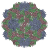
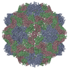
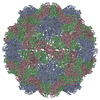
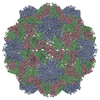
| Title | Three Dimensional Structure of Physalis Mottle Virus : Implications for the Viral Assembly | ||||||
 Components Components | COAT PROTEIN | ||||||
 Keywords Keywords | VIRUS / COAT PROTEIN (VIRAL) / ICOSAHEDRAL VIRUS / CAPSID PROTEIN / VIRION | ||||||
| Function / homology |  Function and homology information Function and homology information | ||||||
| Biological species |  PHYSALIS MOTTLE VIRUS PHYSALIS MOTTLE VIRUS | ||||||
| Method |  X-RAY DIFFRACTION / MOLECULAR REPLACEMENT / Resolution: 3.8 Å X-RAY DIFFRACTION / MOLECULAR REPLACEMENT / Resolution: 3.8 Å | ||||||
 Authors Authors | Krishna, S.S. / Hiremath, C.N. / Munshi, S.K. / Prahadeeswaran, D. / Sastri, M. / Savithri, H.S. / Murthy, M.R.N. | ||||||
 Citation Citation | Journal: J.Mol.Biol. / Year: 1999 Title: Three Dimensional Structure of Physalis Movirus: Implications for the Viral Assembly Authors: Krishna, S.S. / Hiremath, C.N. / Munshi, S.K. / Prahadeeswaran, D. / Sastri, M. / Savithri, H.S. / Murthy, M.R.N. #1: Journal: J.Mol.Biol. / Year: 1999 Title: Identification of a Discrete Intermediate in the Assembly/Disassembly of Physalis Mottle Tymovirus Through Mutational Analysis Authors: Sastri, M. / Reddy, S. / Krishna, S.S. / Murthy, M.R.N. / Savithri, H.S. #2: Journal: J.Mol.Biol. / Year: 1997 Title: Assembly of Physalis Mottle Virus Capsid Protein in Escherichia Coli and the Role of Amino and Carboxy Termini in the Formation of the Icosahedral Particles Authors: Sastri, M. / Kekuda, R. / Gopinath, K. / Kumar, C.T.R. / Jagath, J.R. / Savithri, H.S. #3: Journal: Virology / Year: 1993 Title: Architecture of Physalis Mottle Tymovirus as Probed by Monoclonal Antibodies and Cross-Linking Studies Authors: Kekuda, R. / Karande, A.A. / Jacob, A.N.K. / Savithri, H.S. #4: Journal: Acta Crystallogr.,Sect.B / Year: 1990Title: Structure of Belladonna Mottle Virus: Cross Rotation Function Studies with Southern Bean Mosaic Virus Authors: Hiremath, C.N. / Munshi, S.K. / Murthy, M.R.N. #5: Journal: J.Biol.Chem. / Year: 1989 Title: Primary Structure of Belladonna Mottle Virus Coat Protein Authors: Suryanarayana, S. / Rao, N.A. / Murthy, M.R.N. / Savithri, H.S. #6: Journal: J.Gen.Virol. / Year: 1987Title: Stability of Belladonna Mottle Virus Particles: The Role of Polyamines and Calcium Authors: Savithri, H.S. / Munshi, S.K. / Suryanarayana, S. / Divakar, S. / Murthy, M.R.N. #7: Journal: Acta Crystallogr.,Sect.B / Year: 1987Title: Symmetry of Belladonna Mottle Virus: Rotation Function Studies Authors: Munshi, S.K. / Hiremath, C.N. / Murthy, M.R.N. / Savithri, H.S. | ||||||
| History |
| ||||||
| Remark 700 | SHEET THE SHEET STRUCTURE OF THIS MOLECULE IS BIFURCATED. IN ORDER TO REPRESENT THIS FEATURE IN ... SHEET THE SHEET STRUCTURE OF THIS MOLECULE IS BIFURCATED. IN ORDER TO REPRESENT THIS FEATURE IN THE SHEET RECORDS BELOW, TWO SHEETS ARE DEFINED. |
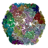
- Structure visualization
| Structure viewer | Molecule: MolmilJmol/JSmol |
|---|
- Downloads & links
-Download
| PDBx/mmCIF format | 1qjz.cif.gz | 100.7 KB | Display | PDBx/mmCIF format |
|---|---|---|---|---|
| PDB format | pdb1qjz.ent.gz | 75.3 KB | Display | PDB format |
| PDBx/mmJSON format | 1qjz.json.gz | Tree view | PDBx/mmJSON format | |
| Others |  Other downloads Other downloads |
-Validation report
| Arichive directory | https://data.pdbj.org/pub/pdb/validation_reports/qj/1qjzftp://data.pdbj.org/pub/pdb/validation_reports/qj/1qjz | HTTPS FTP |
|---|
-Related structure data
| Related structure data |  1auyS S: Starting model for refinement |
|---|---|
| Similar structure data |


-Links
 PDBj
PDBj
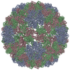
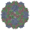
- Assembly
| Deposited unit | 
| ||||||||
|---|---|---|---|---|---|---|---|---|---|
| 1 | x 60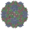
| ||||||||
| 2 |
| ||||||||
| 3 | x 5
| ||||||||
| 4 | x 6
| ||||||||
| 5 | 
| ||||||||
| Unit cell |
| ||||||||
| Symmetry | Point symmetry: (Schoenflies symbol: I (icosahedral)) |
-Components
| #1: Protein | Mass: 19989.924 Da / Num. of mol.: 3 / Source method: isolated from a natural source / Source: (natural) PHYSALIS MOTTLE VIRUS / Strain: IOWA / References: UniProt: P36351 |
|---|
-Experimental details
-Experiment
| Experiment | Method: X-RAY DIFFRACTION / Number of used crystals: 48 |
|---|
- Sample preparation
| Crystal grow | pH: 5.6 / Details: pH 5.60 | ||||||||||||||||||||||||||||||||||||
|---|---|---|---|---|---|---|---|---|---|---|---|---|---|---|---|---|---|---|---|---|---|---|---|---|---|---|---|---|---|---|---|---|---|---|---|---|---|
| Crystal grow | *PLUS Method: vapor diffusion | ||||||||||||||||||||||||||||||||||||
| Components of the solutions | *PLUS
|
-Data collection
| Diffraction | Mean temperature: 295 K |
|---|---|
| Diffraction source | Source: ROTATING ANODE / Type: ENRAF-NONIUS / Wavelength: 1.5418 |
| Detector | Detector: OSCILLATION CAMERA / Details: MIRRORS |
| Radiation | Protocol: SINGLE WAVELENGTH / Monochromatic (M) / Laue (L): M / Scattering type: x-ray |
| Radiation wavelength | Wavelength: 1.5418 Å / Relative weight: 1 |
| Reflection | Resolution: 3.8→60 Å / Num. obs: 137259 / % possible obs: 40 % / Observed criterion σ(I): 2 |
- Processing
| Software |
| ||||||||||||||||||||||||||||||||||||||||||||||||||||||||||||
|---|---|---|---|---|---|---|---|---|---|---|---|---|---|---|---|---|---|---|---|---|---|---|---|---|---|---|---|---|---|---|---|---|---|---|---|---|---|---|---|---|---|---|---|---|---|---|---|---|---|---|---|---|---|---|---|---|---|---|---|---|---|
| Refinement | Method to determine structure: MOLECULAR REPLACEMENT Starting model: PDB ENTRY 1AUY Resolution: 3.8→10 Å / Data cutoff high absF: 10000000 / Data cutoff low absF: 0.001 / σ(F): 2
| ||||||||||||||||||||||||||||||||||||||||||||||||||||||||||||
| Refinement step | Cycle: LAST / Resolution: 3.8→10 Å
| ||||||||||||||||||||||||||||||||||||||||||||||||||||||||||||
| Refine LS restraints |
| ||||||||||||||||||||||||||||||||||||||||||||||||||||||||||||
| Refine LS restraints NCS | NCS model details: STRICT | ||||||||||||||||||||||||||||||||||||||||||||||||||||||||||||
| Xplor file | Serial no: 1 / Param file: PARHCSDX.PRO / Topol file: TOPHCSDX.PRO | ||||||||||||||||||||||||||||||||||||||||||||||||||||||||||||
| Software | *PLUS Version: 3.1 / Classification: refinement | ||||||||||||||||||||||||||||||||||||||||||||||||||||||||||||
| Refinement | *PLUS | ||||||||||||||||||||||||||||||||||||||||||||||||||||||||||||
| Solvent computation | *PLUS | ||||||||||||||||||||||||||||||||||||||||||||||||||||||||||||
| Displacement parameters | *PLUS | ||||||||||||||||||||||||||||||||||||||||||||||||||||||||||||
| Refine LS restraints | *PLUS
| ||||||||||||||||||||||||||||||||||||||||||||||||||||||||||||
| LS refinement shell | *PLUS Highest resolution: 3.8 Å / Lowest resolution: 3.96 Å / Rfactor Rwork: 0.42 / Num. reflection obs: 5434 |
