 Movie
Movie Controller


+ Open data

- Basic information
| Entry | Database: PDB / ID: 1.0E+57 | ||||||
|---|---|---|---|---|---|---|---|
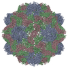
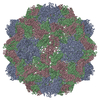
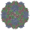
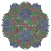
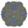
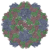
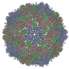
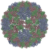
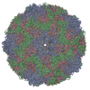
| Title | PHYSALIS MOTTLE VIRUS: EMPTY CAPSID | ||||||
 Components Components | PHYSALIS MOTTLE VIRUS | ||||||
 Keywords Keywords | VIRUS / COAT PROTEIN (VIRAL) / ICOSAHEDRAL VIRUS | ||||||
| Function / homology |  Function and homology information Function and homology information | ||||||
| Biological species |  PHYSALIS MOTTLE VIRUS PHYSALIS MOTTLE VIRUS | ||||||
| Method |  X-RAY DIFFRACTION / MOLECULAR REPLACEMENT / Resolution: 3.2 Å X-RAY DIFFRACTION / MOLECULAR REPLACEMENT / Resolution: 3.2 Å | ||||||
 Authors Authors | Krishna, S.S. / Sastri, M. / Savithri, H.S. / Murthy, M.R.N. | ||||||
 Citation Citation | Journal: J.Mol.Biol. / Year: 2001 Title: Structural Studies on the Empty Capsids of Physalis Mottle Virus Authors: Krishna, S.S. / Sastri, M. / Savithri, H.S. / Murthy, M.R.N. #1: Journal: J.Mol.Biol. / Year: 1999Title: Three Dimensional Structure of Physalis Movirus: Implications for the Viral Assembly Authors: Krishna, S.S. / Hiremath, C.N. / Munshi, S.K. / Prahadeeswaran, D. / Sastri, M. / Savithri, H.S. / Murthy, M.R.N. #2: Journal: J.Mol.Biol. / Year: 1999 Title: Identification of a Discrete Intermediate in the Assembly/Disassembly of Physalis Mottle Tymovirus Through Mutational Analysis Authors: Sastri, M. / Reddy, S. / Krishna, S.S. / Murthy, M.R.N. / Savithri, H.S. #3: Journal: J.Mol.Biol. / Year: 1997 Title: Assembly of Physalis Mottle Virus Capsid Protein in Escherichia Coli and the Role of Amino and Carboxy Termini in the Formation of the Icosahedral Particles Authors: Sastri, M. / Kekuda, R. / Gopinath, K. / Kumar, C.T.R. / Jagath, J.R. / Savithri, H.S. #4: Journal: Virology / Year: 1993 Title: Architecture of Physalis Mottle Tymovirus as Probed by Monoclonal Antibodies and Cross-Linking Studies Authors: Kekuda, R. / Karande, A.A. / Jacob, A.N.K. / Savithri, H.S. #5: Journal: Acta Crystallogr.,Sect.B / Year: 1990Title: Structure of Belladonna Mottle Virus: Cross-Rotation Function Studies with Southern Bean Mosaic Virus Authors: Hiremath, C.N. / Munshi, S.K. / Murthy, M.R.N. #6: Journal: J.Biol.Chem. / Year: 1989 Title: Primary Structure of Belladonna Mottle Virus Coat Protein Authors: Suryanarayana, S. / Rao, N.A. / Murthy, M.R.N. / Savithri, H.S. #7: Journal: J.Gen.Virol. / Year: 1987Title: Stability of Belladonna Mottle Virus Particles: The Role of Polyamines and Calcium Authors: Savithri, H.S. / Munshi, S.K. / Suryanarayana, S. / Divakar, S. / Murthy, M.R.N. #8: Journal: Acta Crystallogr.,Sect.B / Year: 1987Title: Symmetry of Belladonna Mottle Virus: Rotation Function Studies Authors: Munshi, S.K. / Hiremath, C.N. / Murthy, M.R.N. / Savithri, H.S. | ||||||
| History |
|
- Structure visualization
| Structure viewer | Molecule: MolmilJmol/JSmol |
|---|
- Downloads & links
-Download
| PDBx/mmCIF format | 1e57.cif.gz | 95.2 KB | Display | PDBx/mmCIF format |
|---|---|---|---|---|
| PDB format | pdb1e57.ent.gz | 70.3 KB | Display | PDB format |
| PDBx/mmJSON format | 1e57.json.gz | Tree view | PDBx/mmJSON format | |
| Others |  Other downloads Other downloads |
-Validation report
| Arichive directory | https://data.pdbj.org/pub/pdb/validation_reports/e5/1e57ftp://data.pdbj.org/pub/pdb/validation_reports/e5/1e57 | HTTPS FTP |
|---|
-Related structure data
| Related structure data |  1qjzS S: Starting model for refinement |
|---|---|
| Similar structure data |

-Links
 PDBj
PDBj
- Assembly
| Deposited unit | 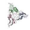
| ||||||||
|---|---|---|---|---|---|---|---|---|---|
| 1 | x 60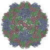
| ||||||||
| 2 |
| ||||||||
| 3 | x 5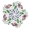
| ||||||||
| 4 | x 6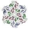
| ||||||||
| 5 | 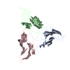
| ||||||||
| 6 | x 60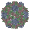
| ||||||||
| Unit cell |
| ||||||||
| Symmetry | Point symmetry: (Schoenflies symbol: I (icosahedral)) |
-Components
| #1: Protein | Mass: 19989.924 Da / Num. of mol.: 3 / Fragment: EMPTY CAPSID Source method: isolated from a genetically manipulated source Source: (gene. exp.) PHYSALIS MOTTLE VIRUS / Plasmid: PET3D / Production host:  |
|---|
-Experimental details
-Experiment
| Experiment | Method: X-RAY DIFFRACTION / Number of used crystals: 1 |
|---|
- Sample preparation
| Crystal grow | pH: 5.6 Details: PH 5.6 SODIUM ACETATE; WITH 20% PEG AS PRECIPITANT. | ||||||||||||||||||||||||||||||||||||
|---|---|---|---|---|---|---|---|---|---|---|---|---|---|---|---|---|---|---|---|---|---|---|---|---|---|---|---|---|---|---|---|---|---|---|---|---|---|
| Crystal grow | *PLUS Method: unknown | ||||||||||||||||||||||||||||||||||||
| Components of the solutions | *PLUS
|
-Data collection
| Diffraction | Mean temperature: 295 K |
|---|---|
| Diffraction source | Source: ROTATING ANODE / Type: RIGAKU / Wavelength: 1.5418 |
| Detector | Type: MARRESEARCH / Detector: IMAGE PLATE / Details: MIRRORS |
| Radiation | Protocol: SINGLE WAVELENGTH / Monochromatic (M) / Laue (L): M / Scattering type: x-ray |
| Radiation wavelength | Wavelength: 1.5418 Å / Relative weight: 1 |
| Reflection | Resolution: 3.2→30 Å / Num. obs: 605968 / % possible obs: 36 % / Observed criterion σ(I): 2 / Rmerge(I) obs: 0.149 |
- Processing
| Software |
| ||||||||||||||||||||||||||||||||||||||||||||||||||||||||||||
|---|---|---|---|---|---|---|---|---|---|---|---|---|---|---|---|---|---|---|---|---|---|---|---|---|---|---|---|---|---|---|---|---|---|---|---|---|---|---|---|---|---|---|---|---|---|---|---|---|---|---|---|---|---|---|---|---|---|---|---|---|---|
| Refinement | Method to determine structure: MOLECULAR REPLACEMENT Starting model: PDB ENTRY 1QJZ Resolution: 3.2→10 Å / Data cutoff high absF: 10000000 / σ(F): 4
| ||||||||||||||||||||||||||||||||||||||||||||||||||||||||||||
| Refinement step | Cycle: LAST / Resolution: 3.2→10 Å
| ||||||||||||||||||||||||||||||||||||||||||||||||||||||||||||
| Refine LS restraints |
| ||||||||||||||||||||||||||||||||||||||||||||||||||||||||||||
| Refine LS restraints NCS | NCS model details: STRICT | ||||||||||||||||||||||||||||||||||||||||||||||||||||||||||||
| Software | *PLUS Name: CNS / Version: 0.4 / Classification: refinement | ||||||||||||||||||||||||||||||||||||||||||||||||||||||||||||
| Refine LS restraints | *PLUS
|
