 Movie
Movie Controller

[English] 日本語
 Yorodumi
- PDB-2fyc: Crystal structure of the catalytic domain of bovine beta1,4-galac... -
+ Open data

- Basic information
| Entry | Database: PDB / ID: 2fyc | ||||||
|---|---|---|---|---|---|---|---|
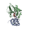
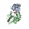
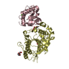
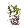
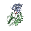
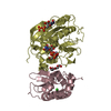
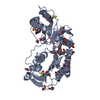
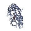
| Title | Crystal structure of the catalytic domain of bovine beta1,4-galactosyltransferase-I in complex with alpha-lactalbumin, Ca and UDP-galactose | ||||||
 Components Components |
| ||||||
 Keywords Keywords | TRANSFERASE / lactose synthase / M344H mutation / Ca binding | ||||||
| Function / homology |  Function and homology information Function and homology informationLactose synthesis / Keratan sulfate biosynthesis / Interaction With Cumulus Cells And The Zona Pellucida / Lactose synthesis / N-Glycan antennae elongation / carbohydrate derivative biosynthetic process / lactose synthase / neolactotriaosylceramide beta-1,4-galactosyltransferase / beta-N-acetylglucosaminylglycopeptide beta-1,4-galactosyltransferase / N-acetyllactosamine synthase ...Lactose synthesis / Keratan sulfate biosynthesis / Interaction With Cumulus Cells And The Zona Pellucida / Lactose synthesis / N-Glycan antennae elongation / carbohydrate derivative biosynthetic process / lactose synthase / neolactotriaosylceramide beta-1,4-galactosyltransferase / beta-N-acetylglucosaminylglycopeptide beta-1,4-galactosyltransferase / N-acetyllactosamine synthase / N-acetyllactosamine synthase activity / positive regulation of circulating fibrinogen levels / beta-N-acetylglucosaminylglycopeptide beta-1,4-galactosyltransferase activity / penetration of zona pellucida / UDP-galactosyltransferase activity / regulation of acrosome reaction / Golgi trans cisterna / lactose synthase activity / lactose biosynthetic process / macrophage migration / oligosaccharide biosynthetic process / development of animal secondary sexual characteristics / desmosome / acute inflammatory response / galactose metabolic process / positive regulation of epithelial cell proliferation involved in wound healing / binding of sperm to zona pellucida / protein N-linked glycosylation / angiogenesis involved in wound healing / Neutrophil degranulation / Transferases; Glycosyltransferases; Hexosyltransferases / Golgi cisterna membrane / epithelial cell development / alpha-tubulin binding / beta-tubulin binding / extracellular matrix organization / epithelial cell proliferation / lipid metabolic process / filopodium / brush border membrane / negative regulation of epithelial cell proliferation / lysozyme activity / manganese ion binding / basolateral plasma membrane / defense response to Gram-negative bacterium / cell adhesion / defense response to Gram-positive bacterium / positive regulation of apoptotic process / external side of plasma membrane / calcium ion binding / Golgi apparatus / protein-containing complex / : / extracellular region / identical protein binding Similarity search - Function | ||||||
| Biological species |  | ||||||
| Method |  X-RAY DIFFRACTION / SYNCHROTRON / MOLECULAR REPLACEMENT / Resolution: 2 Å X-RAY DIFFRACTION / SYNCHROTRON / MOLECULAR REPLACEMENT / Resolution: 2 Å | ||||||
 Authors Authors | Ramakrishnan, B. / Ramasamy, V. / Qasba, P.K. | ||||||
 Citation Citation | Journal: J.Mol.Biol. / Year: 2006 Title: Structural Snapshots of beta-1,4-Galactosyltransferase-I Along the Kinetic Pathway. Authors: Ramakrishnan, B. / Ramasamy, V. / Qasba, P.K. | ||||||
| History |
|
- Structure visualization
| Structure viewer | Molecule: MolmilJmol/JSmol |
|---|
- Downloads & links
-Download
| PDBx/mmCIF format | 2fyc.cif.gz | 196.1 KB | Display | PDBx/mmCIF format |
|---|---|---|---|---|
| PDB format | pdb2fyc.ent.gz | 151.9 KB | Display | PDB format |
| PDBx/mmJSON format | 2fyc.json.gz | Tree view | PDBx/mmJSON format | |
| Others |  Other downloads Other downloads |
-Validation report
| Arichive directory | https://data.pdbj.org/pub/pdb/validation_reports/fy/2fycftp://data.pdbj.org/pub/pdb/validation_reports/fy/2fyc | HTTPS FTP |
|---|
-Related structure data
| Related structure data |  2fy7C  2fyaC  2fybC  2fydC  1oqmS C: citing same article ( S: Starting model for refinement |
|---|---|
| Similar structure data |


-Links
 PDBj
PDBj


- Assembly
| Deposited unit | 
| ||||||||
|---|---|---|---|---|---|---|---|---|---|
| 1 | 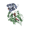
| ||||||||
| 2 | 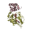
| ||||||||
| Unit cell |
| ||||||||
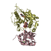
| Details | 1:1 complex between the alpha-lactalbumin and the catalytic domain of beta 1, 4-galactosyltransferase |
-Components
-Protein , 2 types, 4 molecules ACBD
| #1: Protein | Mass: 14015.835 Da / Num. of mol.: 2 / Mutation: C342T, M344H Source method: isolated from a genetically manipulated source Source: (gene. exp.)  #2: Protein | Mass: 32856.531 Da / Num. of mol.: 2 / Fragment: residues 57-329 Source method: isolated from a genetically manipulated source Source: (gene. exp.) References: UniProt: P08037, Transferases; Glycosyltransferases; Hexosyltransferases |
|---|
-Non-polymers , 5 types, 774 molecules 

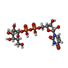


| #3: Chemical | ChemComp-CA /  Mass: 40.078 Da / Num. of mol.: 4 / Source method: obtained synthetically / Formula: Ca Mass: 40.078 Da / Num. of mol.: 4 / Source method: obtained synthetically / Formula: Ca#4: Chemical | ChemComp-MES / |  Mass: 195.237 Da / Num. of mol.: 1 / Source method: obtained synthetically / Formula: C6H13NO4S / Comment: pH buffer*YM Mass: 195.237 Da / Num. of mol.: 1 / Source method: obtained synthetically / Formula: C6H13NO4S / Comment: pH buffer*YM#5: Chemical |  Mass: 566.302 Da / Num. of mol.: 2 / Source method: obtained synthetically / Formula: C15H24N2O17P2 Mass: 566.302 Da / Num. of mol.: 2 / Source method: obtained synthetically / Formula: C15H24N2O17P2#6: Chemical |  Type: RNA linking / Mass: 404.161 Da / Num. of mol.: 2 / Source method: obtained synthetically / Formula: C9H14N2O12P2 / Comment: UDP*YM Type: RNA linking / Mass: 404.161 Da / Num. of mol.: 2 / Source method: obtained synthetically / Formula: C9H14N2O12P2 / Comment: UDP*YM#7: Water | ChemComp-HOH / | Mass: 18.015 Da / Num. of mol.: 765 / Source method: isolated from a natural source / Formula: H2O |
|---|
-Details
| Has protein modification | Y |
|---|
-Experimental details
-Experiment
| Experiment | Method: X-RAY DIFFRACTION / Number of used crystals: 1 |
|---|
- Sample preparation
| Crystal | Density Matthews: 2.82 Å3/Da / Density % sol: 56.41 % |
|---|---|
| Crystal grow | Temperature: 298 K / Method: vapor diffusion / pH: 6.5 Details: 500 Mes-NaOH, 6% PEG 4000%, pH 6.5, VAPOR DIFFUSION, temperature 298K |
-Data collection
| Diffraction | Mean temperature: 100 K |
|---|---|
| Diffraction source | Source: SYNCHROTRON / Site: NSLS  / Beamline: X9B / Wavelength: 0.98 Å / Beamline: X9B / Wavelength: 0.98 Å |
| Detector | Type: ADSC QUANTUM 4 / Detector: CCD / Date: Jun 13, 2003 / Details: mirrors |
| Radiation | Monochromator: graphite / Protocol: SINGLE WAVELENGTH / Monochromatic (M) / Laue (L): M / Scattering type: x-ray |
| Radiation wavelength | Wavelength: 0.98 Å / Relative weight: 1 |
| Reflection | Resolution: 1.98→32.56 Å / Num. obs: 69858 / % possible obs: 96 % / Observed criterion σ(I): 1 / Redundancy: 2.8 % / Biso Wilson estimate: 19.3 Å2 / Rsym value: 0.043 / Net I/σ(I): 17 |
| Reflection shell | Resolution: 1.98→2.05 Å / Redundancy: 2.3 % / Mean I/σ(I) obs: 1.8 / Num. unique all: 6450 / Rsym value: 0.465 / % possible all: 89 |
- Processing
| Software |
| ||||||||||||||||||||||||||||||||||||||||||||||||||||||||||||||||||||||||||||||||
|---|---|---|---|---|---|---|---|---|---|---|---|---|---|---|---|---|---|---|---|---|---|---|---|---|---|---|---|---|---|---|---|---|---|---|---|---|---|---|---|---|---|---|---|---|---|---|---|---|---|---|---|---|---|---|---|---|---|---|---|---|---|---|---|---|---|---|---|---|---|---|---|---|---|---|---|---|---|---|---|---|---|
| Refinement | Method to determine structure: MOLECULAR REPLACEMENT Starting model: PDB ENTRY 1OQM Resolution: 2→32.56 Å / Rfactor Rfree error: 0.003 / Data cutoff high absF: 1664884.26 / Data cutoff low absF: 0 / Isotropic thermal model: RESTRAINED / Cross valid method: THROUGHOUT / σ(F): 0 / Stereochemistry target values: Engh & Huber
| ||||||||||||||||||||||||||||||||||||||||||||||||||||||||||||||||||||||||||||||||
| Solvent computation | Solvent model: FLAT MODEL / Bsol: 61.4715 Å2 / ksol: 0.338919 e/Å3 | ||||||||||||||||||||||||||||||||||||||||||||||||||||||||||||||||||||||||||||||||
| Displacement parameters | Biso mean: 36.9 Å2
| ||||||||||||||||||||||||||||||||||||||||||||||||||||||||||||||||||||||||||||||||
| Refine analyze |
| ||||||||||||||||||||||||||||||||||||||||||||||||||||||||||||||||||||||||||||||||
| Refinement step | Cycle: LAST / Resolution: 2→32.56 Å
| ||||||||||||||||||||||||||||||||||||||||||||||||||||||||||||||||||||||||||||||||
| Refine LS restraints |
| ||||||||||||||||||||||||||||||||||||||||||||||||||||||||||||||||||||||||||||||||
| LS refinement shell | Resolution: 2→2.13 Å / Rfactor Rfree error: 0.01 / Total num. of bins used: 6
| ||||||||||||||||||||||||||||||||||||||||||||||||||||||||||||||||||||||||||||||||
| Xplor file |
|
