 Movie
Movie Controller

[English] 日本語
 Yorodumi
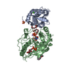
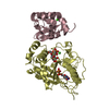
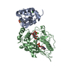
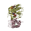
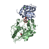
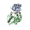
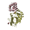
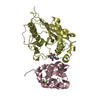
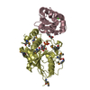
- PDB-1pzy: W314A-BETA1,4-GALACTOSYLTRANSFERASE-I COMPLEXED WITH ALPHA-LACTAL... -
+ Open data

- Basic information
| Entry | Database: PDB / ID: 1pzy | ||||||
|---|---|---|---|---|---|---|---|
| Title | W314A-BETA1,4-GALACTOSYLTRANSFERASE-I COMPLEXED WITH ALPHA-LACTALBUMIN IN THE PRESENCE OF N-ACETYLGLUCOSAMINE, UDP AND MANGANESE | ||||||
 Components Components |
| ||||||
 Keywords Keywords | TRANSFERASE ACTIVATOR/TRANSFERASE / Beta1 / 4-Galactosyltransferase-I Tryptophan Mutant / Flexible loop Conformation / Protease Digetion / Substrate Binding / Catalytic Mechanism / TRANSFERASE ACTIVATOR-TRANSFERASE COMPLEX | ||||||
| Function / homology |  Function and homology information Function and homology informationLactose synthesis / Keratan sulfate biosynthesis / Interaction With Cumulus Cells And The Zona Pellucida / Lactose synthesis / N-Glycan antennae elongation / carbohydrate derivative biosynthetic process / lactose synthase / neolactotriaosylceramide beta-1,4-galactosyltransferase / beta-N-acetylglucosaminylglycopeptide beta-1,4-galactosyltransferase / N-acetyllactosamine synthase ...Lactose synthesis / Keratan sulfate biosynthesis / Interaction With Cumulus Cells And The Zona Pellucida / Lactose synthesis / N-Glycan antennae elongation / carbohydrate derivative biosynthetic process / lactose synthase / neolactotriaosylceramide beta-1,4-galactosyltransferase / beta-N-acetylglucosaminylglycopeptide beta-1,4-galactosyltransferase / N-acetyllactosamine synthase / N-acetyllactosamine synthase activity / positive regulation of circulating fibrinogen levels / beta-N-acetylglucosaminylglycopeptide beta-1,4-galactosyltransferase activity / penetration of zona pellucida / UDP-galactosyltransferase activity / regulation of acrosome reaction / Golgi trans cisterna / lactose synthase activity / lactose biosynthetic process / macrophage migration / oligosaccharide biosynthetic process / development of animal secondary sexual characteristics / desmosome / acute inflammatory response / galactose metabolic process / positive regulation of epithelial cell proliferation involved in wound healing / binding of sperm to zona pellucida / protein N-linked glycosylation / angiogenesis involved in wound healing / Neutrophil degranulation / Transferases; Glycosyltransferases; Hexosyltransferases / Golgi cisterna membrane / epithelial cell development / alpha-tubulin binding / beta-tubulin binding / extracellular matrix organization / epithelial cell proliferation / filopodium / lipid metabolic process / brush border membrane / negative regulation of epithelial cell proliferation / lysozyme activity / manganese ion binding / defense response to Gram-negative bacterium / basolateral plasma membrane / cell adhesion / defense response to Gram-positive bacterium / positive regulation of apoptotic process / external side of plasma membrane / calcium ion binding / Golgi apparatus / protein-containing complex / : / extracellular region / identical protein binding Similarity search - Function | ||||||
| Biological species |  | ||||||
| Method |  X-RAY DIFFRACTION / MOLECULAR REPLACEMENT / Resolution: 2.3 Å X-RAY DIFFRACTION / MOLECULAR REPLACEMENT / Resolution: 2.3 Å | ||||||
 Authors Authors | Ramasamy, V. / Ramakrishnan, B. / Boeggeman, E. / Qasba, P.K. | ||||||
 Citation Citation | Journal: J.Mol.Biol. / Year: 2003 Title: The Role of Tryptophan 314 in the Conformational Changes of beta 1,4-Galactosyltransferase-I Authors: Ramasamy, V. / Ramakrishnan, B. / Boeggeman, E. / Qasba, P.K. | ||||||
| History |
|
- Structure visualization
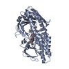
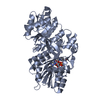
| Structure viewer | Molecule: MolmilJmol/JSmol |
|---|
- Downloads & links
-Download
| PDBx/mmCIF format | 1pzy.cif.gz | 178 KB | Display | PDBx/mmCIF format |
|---|---|---|---|---|
| PDB format | pdb1pzy.ent.gz | 139.9 KB | Display | PDB format |
| PDBx/mmJSON format | 1pzy.json.gz | Tree view | PDBx/mmJSON format | |
| Others |  Other downloads Other downloads |
-Validation report
| Arichive directory | https://data.pdbj.org/pub/pdb/validation_reports/pz/1pzyftp://data.pdbj.org/pub/pdb/validation_reports/pz/1pzy | HTTPS FTP |
|---|
-Related structure data
| Related structure data |  1pztC  1nmmS S: Starting model for refinement C: citing same article ( |
|---|---|
| Similar structure data |

-Links
 PDBj
PDBj


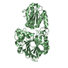
- Assembly
| Deposited unit | 
| ||||||||||
|---|---|---|---|---|---|---|---|---|---|---|---|
| 1 | 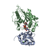
| ||||||||||
| 2 | 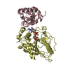
| ||||||||||
| Unit cell |
| ||||||||||
| Details | CHAINS A AND B FORM FIRST, C AND D FORMS SECOND LACTOSE SYNTHASE COMPLEX |
-Components
-Protein , 2 types, 4 molecules ACBD
| #1: Protein | Mass: 14015.835 Da / Num. of mol.: 2 / Fragment: REGULATORY SUBUNIT OF LACTOSE SYNTHASE Source method: isolated from a genetically manipulated source Details: CHAINS A AND B FORM FIRST, B AND D SECOND LACTOSE SYNTHASE COMPLEX Source: (gene. exp.)  #2: Protein | Mass: 32734.449 Da / Num. of mol.: 2 / Fragment: Catalytic Domain, residues 130-402 / Mutation: W314A, C342T Source method: isolated from a genetically manipulated source Details: CHAINS C AND D FORM FIRST, B AND D SECOND LACTOSE SYNTHASE COMPLEX Source: (gene. exp.) Description: N-TERMINAL CARRIES A TAG CONTAINING 13 AMINO ACIDS Plasmid: pET23a / Species (production host): Escherichia coli / Production host: References: UniProt: P08037, lactose synthase, N-acetyllactosamine synthase, beta-N-acetylglucosaminylglycopeptide beta-1,4-galactosyltransferase |
|---|
-Sugars , 1 types, 2 molecules 
| #4: Sugar |  Type: D-saccharide, beta linking / Mass: 221.208 Da / Num. of mol.: 2 Type: D-saccharide, beta linking / Mass: 221.208 Da / Num. of mol.: 2Source method: isolated from a genetically manipulated source Formula: C8H15NO6 |
|---|
-Non-polymers , 4 types, 257 molecules 


| #3: Chemical |  Mass: 40.078 Da / Num. of mol.: 2 / Source method: obtained synthetically / Formula: Ca Mass: 40.078 Da / Num. of mol.: 2 / Source method: obtained synthetically / Formula: Ca#5: Chemical |  Mass: 54.938 Da / Num. of mol.: 2 / Source method: obtained synthetically / Formula: Mn Mass: 54.938 Da / Num. of mol.: 2 / Source method: obtained synthetically / Formula: Mn#6: Chemical |  Type: RNA linking / Mass: 404.161 Da / Num. of mol.: 2 / Source method: obtained synthetically / Formula: C9H14N2O12P2 / Comment: UDP*YM Type: RNA linking / Mass: 404.161 Da / Num. of mol.: 2 / Source method: obtained synthetically / Formula: C9H14N2O12P2 / Comment: UDP*YM#7: Water | ChemComp-HOH / | Mass: 18.015 Da / Num. of mol.: 251 / Source method: isolated from a natural source / Formula: H2O |
|---|
-Details
| Has protein modification | Y |
|---|
-Experimental details
-Experiment
| Experiment | Method: X-RAY DIFFRACTION / Number of used crystals: 1 |
|---|
- Sample preparation
| Crystal | Density Matthews: 2.9 Å3/Da / Density % sol: 57.3 % | ||||||||||||||||||||||||||||||||||||
|---|---|---|---|---|---|---|---|---|---|---|---|---|---|---|---|---|---|---|---|---|---|---|---|---|---|---|---|---|---|---|---|---|---|---|---|---|---|
| Crystal grow | Temperature: 298 K / Method: vapor diffusion, hanging drop / pH: 6.1 Details: 11% PEG 4000, 0.1M Sodium chloride and 0.1M Sodium citrate, pH 6.1, VAPOR DIFFUSION, HANGING DROP, temperature 298K | ||||||||||||||||||||||||||||||||||||
| Crystal grow | *PLUS pH: 8 / Method: vapor diffusion, hanging drop | ||||||||||||||||||||||||||||||||||||
| Components of the solutions | *PLUS
|
-Data collection
| Diffraction | Mean temperature: 100 K |
|---|---|
| Diffraction source | Source: ROTATING ANODE / Type: RIGAKU RU200 / Wavelength: 1.5418 Å |
| Detector | Type: MARRESEARCH / Detector: IMAGE PLATE / Date: Aug 31, 2002 / Details: mirrors |
| Radiation | Monochromator: mirrors / Protocol: SINGLE WAVELENGTH / Monochromatic (M) / Laue (L): M / Scattering type: x-ray |
| Radiation wavelength | Wavelength: 1.5418 Å / Relative weight: 1 |
| Reflection | Resolution: 2.3→25 Å / Num. obs: 46294 / % possible obs: 97 % / Observed criterion σ(I): 1 / Redundancy: 3.5 % / Biso Wilson estimate: 28.9 Å2 / Rsym value: 0.07 / Net I/σ(I): 16.4 |
| Reflection shell | Resolution: 2.3→2.38 Å / Redundancy: 3.4 % / Mean I/σ(I) obs: 2.2 / Num. unique all: 4427 / Rsym value: 0.48 / % possible all: 93.4 |
| Reflection | *PLUS Highest resolution: 2.3 Å / Lowest resolution: 25 Å / Num. obs: 47881 / Num. measured all: 161993 / Rmerge(I) obs: 0.07 |
| Reflection shell | *PLUS % possible obs: 93.4 % / Rmerge(I) obs: 0.48 |
- Processing
| Software |
| ||||||||||||||||||||||||||||||||||||
|---|---|---|---|---|---|---|---|---|---|---|---|---|---|---|---|---|---|---|---|---|---|---|---|---|---|---|---|---|---|---|---|---|---|---|---|---|---|
| Refinement | Method to determine structure: MOLECULAR REPLACEMENT Starting model: 1NMM Resolution: 2.3→19.95 Å / Rfactor Rfree error: 0.004 / Isotropic thermal model: RESTRAINED / Cross valid method: THROUGHOUT / σ(F): 0 / Stereochemistry target values: Engh & Huber
| ||||||||||||||||||||||||||||||||||||
| Solvent computation | Solvent model: FLAT MODEL / Bsol: 33.1219 Å2 / ksol: 0.327069 e/Å3 | ||||||||||||||||||||||||||||||||||||
| Displacement parameters | Biso mean: 42.1 Å2
| ||||||||||||||||||||||||||||||||||||
| Refine analyze | Luzzati coordinate error free: 0.37 Å / Luzzati sigma a free: 0.44 Å | ||||||||||||||||||||||||||||||||||||
| Refinement step | Cycle: LAST / Resolution: 2.3→19.95 Å
| ||||||||||||||||||||||||||||||||||||
| Refine LS restraints |
| ||||||||||||||||||||||||||||||||||||
| LS refinement shell | Resolution: 2.3→2.44 Å / Rfactor Rfree error: 0.013 / Total num. of bins used: 6
| ||||||||||||||||||||||||||||||||||||
| Xplor file |
| ||||||||||||||||||||||||||||||||||||
| Refinement | *PLUS Highest resolution: 2.3 Å / Lowest resolution: 25 Å / Rfactor Rfree: 0.254 | ||||||||||||||||||||||||||||||||||||
| Solvent computation | *PLUS | ||||||||||||||||||||||||||||||||||||
| Displacement parameters | *PLUS | ||||||||||||||||||||||||||||||||||||
| Refine LS restraints | *PLUS
|
