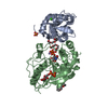
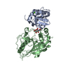
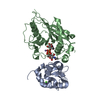
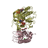
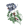
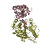
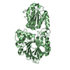
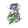
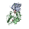
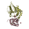
[English] 日本語
Yorodumi
- PDB-1nf5: Crystal Structure of Lactose Synthase, Complex with Glucose -

+
Open data


ID or keywords:

Loading...

-
Basic information

Entry
Database: PDB / ID: 1nf5
TitleCrystal Structure of Lactose Synthase, Complex with Glucose
Components
  • Alpha-lactalbumin
  • beta-1,4-galactosyltransferase
KeywordsTRANSFERASE/TRANSFERASE / LACTOSE SYNHTASE / GLUCOSE BINDING / CONFORMATIONAL CHANGE / transferase modulator / TRANSFERASE-TRANSFERASE COMPLEX
Function / homology
Function and homology information


Lactose synthesis / protein galactosylation / Keratan sulfate biosynthesis / Interaction With Cumulus Cells And The Zona Pellucida / Lactose synthesis / N-Glycan antennae elongation / lactose synthase / neolactotriaosylceramide beta-1,4-galactosyltransferase / beta-N-acetylglucosaminylglycopeptide beta-1,4-galactosyltransferase / N-acetyllactosamine synthase ...Lactose synthesis / protein galactosylation / Keratan sulfate biosynthesis / Interaction With Cumulus Cells And The Zona Pellucida / Lactose synthesis / N-Glycan antennae elongation / lactose synthase / neolactotriaosylceramide beta-1,4-galactosyltransferase / beta-N-acetylglucosaminylglycopeptide beta-1,4-galactosyltransferase / N-acetyllactosamine synthase / N-acetyllactosamine synthase activity / beta-N-acetylglucosaminylglycopeptide beta-1,4-galactosyltransferase activity / regulation of acrosome reaction / UDP-galactosyltransferase activity / penetration of zona pellucida / Golgi trans cisterna / lactose synthase activity / macrophage migration / lactose biosynthetic process / oligosaccharide biosynthetic process / development of secondary sexual characteristics / desmosome / acute inflammatory response / positive regulation of epithelial cell proliferation involved in wound healing / galactose metabolic process / binding of sperm to zona pellucida / angiogenesis involved in wound healing / protein N-linked glycosylation / Neutrophil degranulation / Transferases; Glycosyltransferases; Hexosyltransferases / Golgi cisterna membrane / beta-tubulin binding / epithelial cell development / alpha-tubulin binding / cytoskeletal protein binding / extracellular matrix organization / filopodium / epithelial cell proliferation / brush border membrane / lipid metabolic process / negative regulation of epithelial cell proliferation / lysozyme activity / manganese ion binding / basolateral plasma membrane / cell adhesion / positive regulation of apoptotic process / external side of plasma membrane / calcium ion binding / Golgi apparatus / protein-containing complex / extracellular space / extracellular region / identical protein binding
Similarity search - Function
Lactalbumin / Beta-1,4-galactosyltransferase / Galactosyltransferase, N-terminal / N-terminal region of glycosyl transferase group 7 / Galactosyltransferase, C-terminal / N-terminal domain of galactosyltransferase / Spore Coat Polysaccharide Biosynthesis Protein SpsA; Chain A / Spore Coat Polysaccharide Biosynthesis Protein SpsA; Chain A / Nucleotide-diphospho-sugar transferases / Lysozyme - #10 ...Lactalbumin / Beta-1,4-galactosyltransferase / Galactosyltransferase, N-terminal / N-terminal region of glycosyl transferase group 7 / Galactosyltransferase, C-terminal / N-terminal domain of galactosyltransferase / Spore Coat Polysaccharide Biosynthesis Protein SpsA; Chain A / Spore Coat Polysaccharide Biosynthesis Protein SpsA; Chain A / Nucleotide-diphospho-sugar transferases / Lysozyme - #10 / Glycoside hydrolase family 22 domain / Glycosyl hydrolases family 22 (GH22) domain signature. / Glycoside hydrolase, family 22 / C-type lysozyme/alpha-lactalbumin family / Glycosyl hydrolases family 22 (GH22) domain profile. / Alpha-lactalbumin / lysozyme C / Lysozyme / Lysozyme-like domain superfamily / Alpha-Beta Complex / Orthogonal Bundle / Mainly Alpha / Alpha Beta
Similarity search - Domain/homology
beta-D-glucopyranose / Beta-1,4-galactosyltransferase 1 / Alpha-lactalbumin
Similarity search - Component
Biological speciesMus musculus (house mouse)
Bos taurus (cattle)
MethodX-RAY DIFFRACTION / SYNCHROTRON / MOLECULAR REPLACEMENT / Resolution: 2 Å
AuthorsRamakrishnan, B. / Qasba, P.K.
CitationJournal: J.Mol.Biol. / Year: 2001
Title: Crystal Structure of Lactose Synthase Reveals a Large Conformational Change in its Catalytic Component, the beta-1,4-galactosyltransferase
Authors: Ramakrishnan, B. / Qasba, P.K.
History
DepositionDec 13, 2002Deposition site: RCSB / Processing site: RCSB
SupersessionDec 25, 2002ID: 1J8W
Revision 1.0Dec 25, 2002Provider: repository / Type: Initial release
Revision 1.1Apr 29, 2008Group: Version format compliance
Revision 1.2Jul 13, 2011Group: Version format compliance
Revision 1.3Jul 29, 2020Group: Data collection / Derived calculations / Structure summary
Category: chem_comp / entity ...chem_comp / entity / pdbx_chem_comp_identifier / pdbx_entity_nonpoly / pdbx_struct_conn_angle / struct_conn / struct_site / struct_site_gen
Item: _chem_comp.name / _chem_comp.type ..._chem_comp.name / _chem_comp.type / _entity.pdbx_description / _pdbx_entity_nonpoly.name / _pdbx_struct_conn_angle.ptnr1_auth_comp_id / _pdbx_struct_conn_angle.ptnr1_auth_seq_id / _pdbx_struct_conn_angle.ptnr1_label_asym_id / _pdbx_struct_conn_angle.ptnr1_label_atom_id / _pdbx_struct_conn_angle.ptnr1_label_comp_id / _pdbx_struct_conn_angle.ptnr1_label_seq_id / _pdbx_struct_conn_angle.ptnr3_auth_comp_id / _pdbx_struct_conn_angle.ptnr3_auth_seq_id / _pdbx_struct_conn_angle.ptnr3_label_asym_id / _pdbx_struct_conn_angle.ptnr3_label_atom_id / _pdbx_struct_conn_angle.ptnr3_label_comp_id / _pdbx_struct_conn_angle.ptnr3_label_seq_id / _pdbx_struct_conn_angle.value / _struct_conn.pdbx_dist_value / _struct_conn.ptnr1_auth_comp_id / _struct_conn.ptnr1_auth_seq_id / _struct_conn.ptnr1_label_asym_id / _struct_conn.ptnr1_label_atom_id / _struct_conn.ptnr1_label_comp_id / _struct_conn.ptnr1_label_seq_id / _struct_conn.ptnr2_auth_comp_id / _struct_conn.ptnr2_auth_seq_id / _struct_conn.ptnr2_label_asym_id / _struct_conn.ptnr2_label_atom_id / _struct_conn.ptnr2_label_comp_id / _struct_conn.ptnr2_label_seq_id
Description: Carbohydrate remediation / Provider: repository / Type: Remediation

-
Structure visualization

Structure viewerMolecule:
MolmilJmol/JSmol

Downloads & links

-
Assembly

Deposited unit
A: Alpha-lactalbumin
B: beta-1,4-galactosyltransferase
C: Alpha-lactalbumin
D: beta-1,4-galactosyltransferase
hetero molecules


Theoretical massNumber of molelcules
Total (without water)94,62611
Polymers93,7354
Non-polymers8917
Water10,647591
1
A: Alpha-lactalbumin
B: beta-1,4-galactosyltransferase
hetero molecules


Theoretical massNumber of molelcules
Total (without water)47,4766
Polymers46,8672
Non-polymers6094
Water362
TypeNameSymmetry operationNumber
identity operation1_555x,y,z1
2
C: Alpha-lactalbumin
D: beta-1,4-galactosyltransferase
hetero molecules


Theoretical massNumber of molelcules
Total (without water)47,1505
Polymers46,8672
Non-polymers2823
Water362
TypeNameSymmetry operationNumber
identity operation1_555x,y,z1
Unit cell
Length a, b, c (Å)57.223, 93.898, 99.968
Angle α, β, γ (deg.)90.00, 101.50, 90.00
Int Tables number4
Space group name H-MP1211

-
Components

-
Protein , 2 types, 4 molecules ACBD

#1: Protein Alpha-lactalbumin / / Lactose synthase B protein


Mass: 14015.835 Da / Num. of mol.: 2
Source method: isolated from a genetically manipulated source
Source: (gene. exp.) Mus musculus (house mouse) / Gene: LALBA / Plasmid: pET17a / Species (production host): Escherichia coli / Production host: Escherichia coli BL21 (bacteria) / Strain (production host): BL21 / References: UniProt: P29752
#2: Protein beta-1,4-galactosyltransferase


Mass: 32851.617 Da / Num. of mol.: 2
Source method: isolated from a genetically manipulated source
Source: (gene. exp.) Bos taurus (cattle) / Plasmid: pET23a / Species (production host): Escherichia coli / Production host: Escherichia coli BL21 (bacteria) / Strain (production host): BL21
References: UniProt: P08037, lactose synthase, N-acetyllactosamine synthase, beta-N-acetylglucosaminylglycopeptide beta-1,4-galactosyltransferase

-
Sugars , 1 types, 2 molecules

#5: Sugar ChemComp-BGC / beta-D-glucopyranose / Glucose


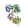
Type: D-saccharide, beta linking / Mass: 180.156 Da / Num. of mol.: 2
Source method: isolated from a genetically manipulated source
Formula: C6H12O6
IdentifierTypeProgram
DGlcpbCONDENSED IUPAC CARBOHYDRATE SYMBOLGMML 1.0
b-D-glucopyranoseCOMMON NAMEGMML 1.0
b-D-GlcpIUPAC CARBOHYDRATE SYMBOLPDB-CARE 1.0
GlcSNFG CARBOHYDRATE SYMBOLGMML 1.0

-
Non-polymers , 4 types, 596 molecules

#3: Chemical ChemComp-CA / CALCIUM ION


Mass: 40.078 Da / Num. of mol.: 2 / Source method: obtained synthetically / Formula: Ca
#4: Chemical ChemComp-PG4 / TETRAETHYLENE GLYCOL / Polyethylene glycol


Mass: 194.226 Da / Num. of mol.: 2 / Source method: obtained synthetically / Formula: C8H18O5 / Comment: precipitant*YM
#6: Chemical ChemComp-EDO / 1,2-ETHANEDIOL / ETHYLENE GLYCOL / Ethylene glycol


Mass: 62.068 Da / Num. of mol.: 1 / Source method: obtained synthetically / Formula: C2H6O2
#7: Water ChemComp-HOH / water / Water


Mass: 18.015 Da / Num. of mol.: 591 / Source method: isolated from a natural source / Formula: H2O

-
Experimental details

-
Experiment

ExperimentMethod: X-RAY DIFFRACTION / Number of used crystals: 1

-
Sample preparation

CrystalDensity Matthews: 2.66 Å3/Da / Density % sol: 53.44 %
Crystal growTemperature: 298 K / Method: vapor diffusion, hanging drop / pH: 5.5
Details: PEG 4000, SODIUM CITRATE, pH 5.50, VAPOR DIFFUSION, HANGING DROP, temperature 298K
Crystal grow
*PLUS
pH: 5.6
Components of the solutions
*PLUS
IDConc.Common nameCrystal-IDSol-IDChemical formulaDetails
120 mg/mlbeta4Gal-T11drop
210 mg/mlLA1drop
3100 mM1reservoirNaCl
4100 mMsodium citrate1reservoirpH5.6
512.5 %(w/v)PEG40001reservoir
6100 mMGlc1drop

-
Data collection

DiffractionMean temperature: 100 K
Diffraction sourceSource: SYNCHROTRON / Site: NSLS / Beamline: X9B / Wavelength: 0.98 / Wavelength: 0.98 Å
DetectorType: ADSC QUANTUM 4 / Detector: CCD / Date: Aug 8, 2000
RadiationMonochromator: GRAPHITE / Protocol: SINGLE WAVELENGTH / Monochromatic (M) / Laue (L): M / Scattering type: x-ray
Radiation wavelengthWavelength: 0.98 Å / Relative weight: 1
ReflectionResolution: 2→25 Å / Num. obs: 57149 / Observed criterion σ(I): 1 / Redundancy: 2.08 % / Biso Wilson estimate: 17.1 Å2 / Rsym value: 0.089
Reflection shellResolution: 2→2.07 Å / Redundancy: 2.08 % / Mean I/σ(I) obs: 2 / Rsym value: 0.335 / % possible all: 77
Reflection
*PLUS
Highest resolution: 2 Å / Lowest resolution: 25 Å / Num. obs: 57639 / Num. measured all: 119864 / Rmerge(I) obs: 0.089

-
Processing

Software
NameVersionClassification
DENZOdata reduction
SCALEPACKdata scaling
AMoREphasing
CNS1refinement
RefinementMethod to determine structure: MOLECULAR REPLACEMENT / Resolution: 2→24.73 Å / Rfactor Rfree error: 0.004 / Isotropic thermal model: RESTRAINED / Cross valid method: THROUGHOUT / σ(F): 0 / Stereochemistry target values: Engh & Huber
RfactorNum. reflection% reflectionSelection details
Rfree0.241 2893 5.1 %RANDOM
Rwork0.19 ---
obs0.19 57146 81.7 %-
all-57149 --
Solvent computationSolvent model: FLAT MODEL / Bsol: 53.2651 Å2 / ksol: 0.354829 e/Å3
Displacement parametersBiso mean: 30.3 Å2
Baniso -1Baniso -2Baniso -3
1-2.72 Å20 Å21.68 Å2
2--1.42 Å20 Å2
3----4.14 Å2
Refine analyzeLuzzati coordinate error free: 0.28 Å / Luzzati sigma a free: 0.29 Å
Refinement stepCycle: LAST / Resolution: 2→24.73 Å
ProteinNucleic acidLigandSolventTotal
Num. atoms6396 0 50 591 7037
Refine LS restraints
Refine-IDTypeDev idealDev ideal target
X-RAY DIFFRACTIONc_bond_d0.011
X-RAY DIFFRACTIONc_bond_d_na
X-RAY DIFFRACTIONc_bond_d_prot
X-RAY DIFFRACTIONc_angle_d
X-RAY DIFFRACTIONc_angle_d_na
X-RAY DIFFRACTIONc_angle_d_prot
X-RAY DIFFRACTIONc_angle_deg1.7
X-RAY DIFFRACTIONc_angle_deg_na
X-RAY DIFFRACTIONc_angle_deg_prot
X-RAY DIFFRACTIONc_dihedral_angle_d24.6
X-RAY DIFFRACTIONc_dihedral_angle_d_na
X-RAY DIFFRACTIONc_dihedral_angle_d_prot
X-RAY DIFFRACTIONc_improper_angle_d1.21
X-RAY DIFFRACTIONc_improper_angle_d_na
X-RAY DIFFRACTIONc_improper_angle_d_prot
X-RAY DIFFRACTIONc_mcbond_it1.451.5
X-RAY DIFFRACTIONc_mcangle_it2.262
X-RAY DIFFRACTIONc_scbond_it2.212
X-RAY DIFFRACTIONc_scangle_it3.182.5
LS refinement shellResolution: 2→2.13 Å / Rfactor Rfree error: 0.014 / Total num. of bins used: 6
RfactorNum. reflection% reflection
Rfree0.308 481 5.4 %
Rwork0.253 8458 -
obs--77 %
Xplor file
Refine-IDSerial noParam fileTopol file
X-RAY DIFFRACTION1PROTEIN_REP.PARAMPROTEIN.TOP
X-RAY DIFFRACTION2ION.PARAMION.TOP
X-RAY DIFFRACTION3WATER.PARAMWATER.TOP
X-RAY DIFFRACTION4BGC_PG.PARBGC_PG.TOP
Refinement
*PLUS
Num. reflection obs: 51690 / Rfactor Rfree: 0.27 / Rfactor Rwork: 0.24
Solvent computation
*PLUS
Displacement parameters
*PLUS
Refine LS restraints
*PLUS
Refine-IDTypeDev ideal
X-RAY DIFFRACTIONc_bond_d0.006
X-RAY DIFFRACTIONc_angle_deg1.4
X-RAY DIFFRACTIONc_dihedral_angle_d
X-RAY DIFFRACTIONc_dihedral_angle_deg24.6
X-RAY DIFFRACTIONc_improper_angle_d
X-RAY DIFFRACTIONc_improper_angle_deg1.21

+
About Yorodumi

-
News

-
Feb 9, 2022. New format data for meta-information of EMDB entries

New format data for meta-information of EMDB entries

  • Version 3 of the EMDB header file is now the official format.
  • The previous official version 1.9 will be removed from the archive.

Related info.:EMDB header

External links:wwPDB to switch to version 3 of the EMDB data model

-
Aug 12, 2020. Covid-19 info

Covid-19 info

URL: https://pdbj.org/emnavi/covid19.php

New page: Covid-19 featured information page in EM Navigator.

Related info.:Covid-19 info / Mar 5, 2020. Novel coronavirus structure data

+
Mar 5, 2020. Novel coronavirus structure data

Novel coronavirus structure data

Related info.:Yorodumi Speices / Aug 12, 2020. Covid-19 info

External links:COVID-19 featured content - PDBj / Molecule of the Month (242):Coronavirus Proteases

+
Jan 31, 2019. EMDB accession codes are about to change! (news from PDBe EMDB page)

EMDB accession codes are about to change! (news from PDBe EMDB page)

  • The allocation of 4 digits for EMDB accession codes will soon come to an end. Whilst these codes will remain in use, new EMDB accession codes will include an additional digit and will expand incrementally as the available range of codes is exhausted. The current 4-digit format prefixed with “EMD-” (i.e. EMD-XXXX) will advance to a 5-digit format (i.e. EMD-XXXXX), and so on. It is currently estimated that the 4-digit codes will be depleted around Spring 2019, at which point the 5-digit format will come into force.
  • The EM Navigator/Yorodumi systems omit the EMD- prefix.

Related info.:Q: What is EMD? / ID/Accession-code notation in Yorodumi/EM Navigator

External links:EMDB Accession Codes are Changing Soon! / Contact to PDBj

+
Jul 12, 2017. Major update of PDB

Major update of PDB

  • wwPDB released updated PDB data conforming to the new PDBx/mmCIF dictionary.
  • This is a major update changing the version number from 4 to 5, and with Remediation, in which all the entries are updated.
  • In this update, many items about electron microscopy experimental information are reorganized (e.g. em_software).
  • Now, EM Navigator and Yorodumi are based on the updated data.

External links:wwPDB Remediation / Enriched Model Files Conforming to OneDep Data Standards Now Available in the PDB FTP Archive

-
Yorodumi

Thousand views of thousand structures

  • Yorodumi is a browser for structure data from EMDB, PDB, SASBDB, etc.
  • This page is also the successor to EM Navigator detail page, and also detail information page/front-end page for Omokage search.
  • The word "yorodu" (or yorozu) is an old Japanese word meaning "ten thousand". "mi" (miru) is to see.

Related info.:EMDB / PDB / SASBDB / Comparison of 3 databanks / Yorodumi Search / Aug 31, 2016. New EM Navigator & Yorodumi / Yorodumi Papers / Jmol/JSmol / Function and homology information / Changes in new EM Navigator and Yorodumi

Read more