 Movie
Movie Controller

[English] 日本語
 Yorodumi
- PDB-2fy7: Crystal structure of the catalytic domain of the human beta1,4-ga... -
+ Open data

- Basic information
| Entry | Database: PDB / ID: 2fy7 | ||||||
|---|---|---|---|---|---|---|---|
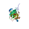
| Title | Crystal structure of the catalytic domain of the human beta1,4-galactosyltransferase mutant M339H in apo form | ||||||
 Components Components | Beta-1,4-galactosyltransferase 1 | ||||||
 Keywords Keywords | TRANSFERASE / M339H mutant / apo enzyme | ||||||
| Function / homology |  Function and homology information Function and homology informationDefective B4GALT1 causes CDG-2d / Interaction With Cumulus Cells And The Zona Pellucida / Defective B4GALT1 causes B4GALT1-CDG (CDG-2d) / Lactose synthesis / galactosyltransferase activity / Keratan sulfate biosynthesis / lactose synthase / neolactotriaosylceramide beta-1,4-galactosyltransferase / beta-N-acetylglucosaminylglycopeptide beta-1,4-galactosyltransferase / N-acetyllactosamine synthase ...Defective B4GALT1 causes CDG-2d / Interaction With Cumulus Cells And The Zona Pellucida / Defective B4GALT1 causes B4GALT1-CDG (CDG-2d) / Lactose synthesis / galactosyltransferase activity / Keratan sulfate biosynthesis / lactose synthase / neolactotriaosylceramide beta-1,4-galactosyltransferase / beta-N-acetylglucosaminylglycopeptide beta-1,4-galactosyltransferase / N-acetyllactosamine synthase / N-acetyllactosamine synthase activity / positive regulation of circulating fibrinogen levels / beta-N-acetylglucosaminylglycopeptide beta-1,4-galactosyltransferase activity / N-Glycan antennae elongation / penetration of zona pellucida / UDP-galactosyltransferase activity / regulation of acrosome reaction / Golgi trans cisterna / lactose synthase activity / lactose biosynthetic process / macrophage migration / oligosaccharide biosynthetic process / development of animal secondary sexual characteristics / desmosome / acute inflammatory response / galactose metabolic process / Pre-NOTCH Processing in Golgi / positive regulation of epithelial cell proliferation involved in wound healing / binding of sperm to zona pellucida / protein N-linked glycosylation / angiogenesis involved in wound healing / Transferases; Glycosyltransferases; Hexosyltransferases / Golgi cisterna membrane / azurophil granule membrane / epithelial cell development / alpha-tubulin binding / beta-tubulin binding / extracellular matrix organization / epithelial cell proliferation / secretory granule membrane / lipid metabolic process / filopodium / brush border membrane / negative regulation of epithelial cell proliferation / manganese ion binding / basolateral plasma membrane / cell adhesion / positive regulation of apoptotic process / Golgi membrane / external side of plasma membrane / Neutrophil degranulation / Golgi apparatus / protein-containing complex / : / extracellular exosome / membrane / identical protein binding / plasma membrane Similarity search - Function | ||||||
| Biological species |  Homo sapiens (human) Homo sapiens (human) | ||||||
| Method |  X-RAY DIFFRACTION / MOLECULAR REPLACEMENT / Resolution: 1.7 Å X-RAY DIFFRACTION / MOLECULAR REPLACEMENT / Resolution: 1.7 Å | ||||||
 Authors Authors | Ramakrishnan, B. / Ramasamy, V. / Qasba, P.K. | ||||||
 Citation Citation | Journal: J.Mol.Biol. / Year: 2006 Title: Structural Snapshots of beta-1,4-Galactosyltransferase-I Along the Kinetic Pathway. Authors: Ramakrishnan, B. / Ramasamy, V. / Qasba, P.K. | ||||||
| History |
|
- Structure visualization
| Structure viewer | Molecule: MolmilJmol/JSmol |
|---|
- Downloads & links
-Download
| PDBx/mmCIF format | 2fy7.cif.gz | 75.6 KB | Display | PDBx/mmCIF format |
|---|---|---|---|---|
| PDB format | pdb2fy7.ent.gz | 55.2 KB | Display | PDB format |
| PDBx/mmJSON format | 2fy7.json.gz | Tree view | PDBx/mmJSON format | |
| Others |  Other downloads Other downloads |
-Validation report
| Arichive directory | https://data.pdbj.org/pub/pdb/validation_reports/fy/2fy7ftp://data.pdbj.org/pub/pdb/validation_reports/fy/2fy7 | HTTPS FTP |
|---|
-Related structure data
| Related structure data |  2fyaC  2fybC  2fycC  2fydC  1oorS C: citing same article ( S: Starting model for refinement |
|---|---|
| Similar structure data |


-Links
 PDBj
PDBj


- Assembly
| Deposited unit | 
| ||||||||
|---|---|---|---|---|---|---|---|---|---|
| 1 |
| ||||||||
| Unit cell |
|
-Components
| #1: Protein | Mass: 32773.230 Da / Num. of mol.: 1 / Fragment: catalytic domain, residues 125-397 / Mutation: R337T, C338T, M340H Source method: isolated from a genetically manipulated source Source: (gene. exp.) Homo sapiens (human) / Gene: B4GALT1, GGTB2 / Plasmid: pET32a / Species (production host): Escherichia coli / Production host:  | ||||
|---|---|---|---|---|---|
| #2: Chemical |   Mass: 150.173 Da / Num. of mol.: 2 / Source method: obtained synthetically / Formula: C6H14O4 Mass: 150.173 Da / Num. of mol.: 2 / Source method: obtained synthetically / Formula: C6H14O4#3: Water | ChemComp-HOH / |  Mass: 18.015 Da / Num. of mol.: 329 / Source method: isolated from a natural source / Formula: H2O Mass: 18.015 Da / Num. of mol.: 329 / Source method: isolated from a natural source / Formula: H2OHas protein modification | Y | |
-Experimental details
-Experiment
| Experiment | Method: X-RAY DIFFRACTION / Number of used crystals: 1 |
|---|
- Sample preparation
| Crystal | Density Matthews: 2.45 Å3/Da / Density % sol: 49.72 % |
|---|---|
| Crystal grow | Temperature: 298 K / Method: vapor diffusion / pH: 5.6 Details: 50 mM sodium citrate buffer, 6% PEG 4000, pH 5.6, VAPOR DIFFUSION, temperature 298K |
-Data collection
| Diffraction | Mean temperature: 100 K |
|---|---|
| Diffraction source | Source: ROTATING ANODE / Type: RIGAKU RU200 / Wavelength: 1.5418 Å |
| Detector | Type: MARRESEARCH / Detector: IMAGE PLATE / Date: Jan 31, 2002 / Details: mirrors |
| Radiation | Monochromator: graphite / Protocol: SINGLE WAVELENGTH / Monochromatic (M) / Laue (L): M / Scattering type: x-ray |
| Radiation wavelength | Wavelength: 1.5418 Å / Relative weight: 1 |
| Reflection | Resolution: 1.6→20 Å / Num. obs: 38674 / % possible obs: 90.3 % / Observed criterion σ(I): 1 / Redundancy: 3.3 % / Biso Wilson estimate: 21.9 Å2 / Rsym value: 0.049 / Net I/σ(I): 15.6 |
| Reflection shell | Resolution: 1.6→1.66 Å / Mean I/σ(I) obs: 1.3 / Num. unique all: 2527 / Rsym value: 0.407 / % possible all: 60 |
- Processing
| Software |
| ||||||||||||||||||||||||||||||||||||||||||||||||||||||||||||||||||||||||||||||||
|---|---|---|---|---|---|---|---|---|---|---|---|---|---|---|---|---|---|---|---|---|---|---|---|---|---|---|---|---|---|---|---|---|---|---|---|---|---|---|---|---|---|---|---|---|---|---|---|---|---|---|---|---|---|---|---|---|---|---|---|---|---|---|---|---|---|---|---|---|---|---|---|---|---|---|---|---|---|---|---|---|---|
| Refinement | Method to determine structure: MOLECULAR REPLACEMENT Starting model: PDB ENTRY 1OOR Resolution: 1.7→19.75 Å / Rfactor Rfree error: 0.004 / Data cutoff high absF: 1637527.64 / Data cutoff low absF: 0 / Isotropic thermal model: RESTRAINED / Cross valid method: THROUGHOUT / σ(F): 0 Details: Residues 343 to 351 could not be traced due to low electron density
| ||||||||||||||||||||||||||||||||||||||||||||||||||||||||||||||||||||||||||||||||
| Solvent computation | Solvent model: FLAT MODEL / Bsol: 43.5119 Å2 / ksol: 0.347019 e/Å3 | ||||||||||||||||||||||||||||||||||||||||||||||||||||||||||||||||||||||||||||||||
| Displacement parameters | Biso mean: 21.2 Å2
| ||||||||||||||||||||||||||||||||||||||||||||||||||||||||||||||||||||||||||||||||
| Refine analyze |
| ||||||||||||||||||||||||||||||||||||||||||||||||||||||||||||||||||||||||||||||||
| Refinement step | Cycle: LAST / Resolution: 1.7→19.75 Å
| ||||||||||||||||||||||||||||||||||||||||||||||||||||||||||||||||||||||||||||||||
| Refine LS restraints |
| ||||||||||||||||||||||||||||||||||||||||||||||||||||||||||||||||||||||||||||||||
| LS refinement shell | Resolution: 1.7→1.81 Å / Rfactor Rfree error: 0.017 / Total num. of bins used: 6
| ||||||||||||||||||||||||||||||||||||||||||||||||||||||||||||||||||||||||||||||||
| Xplor file |
|
