 Movie
Movie Controller

[English] 日本語
 Yorodumi
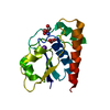
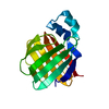
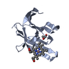
- PDB-2fxi: Arsenate reductase (ArsC from pI258) C10S/C15A double mutant with... -
+ Open data

- Basic information
| Entry | Database: PDB / ID: 2fxi | ||||||
|---|---|---|---|---|---|---|---|
| Title | Arsenate reductase (ArsC from pI258) C10S/C15A double mutant with sulfate in its active site | ||||||
 Components Components | Protein arsC | ||||||
 Keywords Keywords | OXIDOREDUCTASE / HYDROLASE / arsenate reductase / redox enzyme | ||||||
| Function / homology |  Function and homology information Function and homology informationarsenate reductase (thioredoxin) / arsenate reductase (thioredoxin) activity / response to arsenic-containing substance / protein tyrosine phosphatase activity / metal ion binding / cytoplasm Similarity search - Function | ||||||
| Biological species |   Staphylococcus aureus (bacteria) Staphylococcus aureus (bacteria) | ||||||
| Method |  X-RAY DIFFRACTION / SYNCHROTRON / MOLECULAR REPLACEMENT / Resolution: 1.8 Å X-RAY DIFFRACTION / SYNCHROTRON / MOLECULAR REPLACEMENT / Resolution: 1.8 Å | ||||||
 Authors Authors | Loris, R. / Buts, L. / Messens, J. | ||||||
 Citation Citation | Journal: J.Mol.Biol. / Year: 2006 Title: Interplay Between Ion Binding and Catalysis in the Thioredoxin-coupled Arsenate Reductase Family. Authors: Roos, G. / Buts, L. / Van Belle, K. / Brosens, E. / Geerlings, P. / Loris, R. / Wyns, L. / Messens, J. | ||||||
| History |
|
- Structure visualization
| Structure viewer | Molecule: MolmilJmol/JSmol |
|---|
- Downloads & links
-Download
| PDBx/mmCIF format | 2fxi.cif.gz | 43.7 KB | Display | PDBx/mmCIF format |
|---|---|---|---|---|
| PDB format | pdb2fxi.ent.gz | 29.1 KB | Display | PDB format |
| PDBx/mmJSON format | 2fxi.json.gz | Tree view | PDBx/mmJSON format | |
| Others |  Other downloads Other downloads |
-Validation report
| Arichive directory | https://data.pdbj.org/pub/pdb/validation_reports/fx/2fxiftp://data.pdbj.org/pub/pdb/validation_reports/fx/2fxi | HTTPS FTP |
|---|
-Related structure data
| Related structure data |  2cd7C  1ljlS S: Starting model for refinement C: citing same article ( |
|---|---|
| Similar structure data |


-Links
 PDBj
PDBj- Assembly
| Deposited unit | 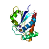
| ||||||||
|---|---|---|---|---|---|---|---|---|---|
| 1 |
| ||||||||
| Unit cell |
|
-Components
| #1: Protein | Mass: 14783.608 Da / Num. of mol.: 1 / Mutation: C10S,C15A Source method: isolated from a genetically manipulated source Source: (gene. exp.) Staphylococcus aureus (bacteria) / Gene: arsC / Plasmid: pI258 / Production host: References: UniProt: P0A006, EC: 1.20.4.-, protein-tyrosine-phosphatase |
|---|---|
| #2: Chemical | ChemComp-K /   Mass: 39.098 Da / Num. of mol.: 1 / Source method: obtained synthetically / Formula: K Mass: 39.098 Da / Num. of mol.: 1 / Source method: obtained synthetically / Formula: K |
| #3: Chemical | ChemComp-SO4 /   Mass: 96.063 Da / Num. of mol.: 1 / Source method: obtained synthetically / Formula: SO4 Mass: 96.063 Da / Num. of mol.: 1 / Source method: obtained synthetically / Formula: SO4 |
| #4: Water | ChemComp-HOH /  Mass: 18.015 Da / Num. of mol.: 168 / Source method: isolated from a natural source / Formula: H2O Mass: 18.015 Da / Num. of mol.: 168 / Source method: isolated from a natural source / Formula: H2O |
-Experimental details
-Experiment
| Experiment | Method: X-RAY DIFFRACTION / Number of used crystals: 1 |
|---|
- Sample preparation
| Crystal | Density Matthews: 1.9 Å3/Da / Density % sol: 35.23 % |
|---|---|
| Crystal grow | Temperature: 293 K / Method: vapor diffusion, hanging drop / pH: 8 Details: 100 mM TRIS ph 8.0, 10 mM DTT, 100 mM KCl, 10 mM K2SO4, 42.5% PEG-4000, VAPOR DIFFUSION, HANGING DROP, temperature 293K |
-Data collection
| Diffraction | Mean temperature: 100 K |
|---|---|
| Diffraction source | Source: SYNCHROTRON / Site: ESRF  / Beamline: ID14-2 / Wavelength: 0.934 Å / Beamline: ID14-2 / Wavelength: 0.934 Å |
| Detector | Type: ADSC QUANTUM 4 / Detector: CCD / Date: Mar 3, 2002 |
| Radiation | Protocol: SINGLE WAVELENGTH / Monochromatic (M) / Laue (L): M / Scattering type: x-ray |
| Radiation wavelength | Wavelength: 0.934 Å / Relative weight: 1 |
| Reflection | Resolution: 1.8→20 Å / Num. all: 10780 / Num. obs: 10780 / % possible obs: 97.9 % / Observed criterion σ(F): 0 / Observed criterion σ(I): 0 / Redundancy: 3.3 % / Biso Wilson estimate: 12.6 Å2 / Rmerge(I) obs: 0.037 / Rsym value: 0.037 / Net I/σ(I): 15.5 |
| Reflection shell | Resolution: 1.8→1.86 Å / Redundancy: 2.3 % / Rmerge(I) obs: 0.08 / Mean I/σ(I) obs: 6.6 / Num. unique all: 914 / Rsym value: 0.08 / % possible all: 83.9 |
- Processing
| Software |
| |||||||||||||||||||||||||
|---|---|---|---|---|---|---|---|---|---|---|---|---|---|---|---|---|---|---|---|---|---|---|---|---|---|---|
| Refinement | Method to determine structure: MOLECULAR REPLACEMENT Starting model: pdb entry 1ljl Resolution: 1.8→20 Å / Cross valid method: THROUGHOUT / σ(F): 0 / σ(I): 0 / Stereochemistry target values: Engh & Huber
| |||||||||||||||||||||||||
| Refinement step | Cycle: LAST / Resolution: 1.8→20 Å
| |||||||||||||||||||||||||
| Refine LS restraints |
|
