 Movie
Movie Controller


+ Open data

- Basic information
| Entry | Database: PDB / ID: 2fjc | ||||||
|---|---|---|---|---|---|---|---|
| Title | Crystal structure of antigen TpF1 from Treponema pallidum | ||||||
 Components Components | Antigen TpF1 | ||||||
 Keywords Keywords | METAL TRANSPORT / Mini ferritin / iron binding protein / antigen | ||||||
| Function / homology |  Function and homology information Function and homology information | ||||||
| Biological species |  Treponema pallidum (bacteria) Treponema pallidum (bacteria) | ||||||
| Method |  X-RAY DIFFRACTION / SYNCHROTRON / MOLECULAR REPLACEMENT / Resolution: 2.5 Å X-RAY DIFFRACTION / SYNCHROTRON / MOLECULAR REPLACEMENT / Resolution: 2.5 Å | ||||||
 Authors Authors | Thumiger, A. / Polenghi, A. / Papinutto, E. / Battistutta, R. / Montecucco, C. / Zanotti, G. | ||||||
 Citation Citation | Journal: Proteins / Year: 2006 Title: Crystal structure of antigen TpF1 from Treponema pallidum. Authors: Thumiger, A. / Polenghi, A. / Papinutto, E. / Battistutta, R. / Montecucco, C. / Zanotti, G. #1: Journal: J.Mol.Biol. / Year: 2002Title: Structure of the neutrophil-activating protein from Helicobacter pylori Authors: Zanotti, G. / Papinutto, E. / Dundon, W. / Battistutta, R. / Seveso, M. / Giudice, G. / Rappuoli, R. / Montecucco, C. #2: Journal: Nat.Struct.Mol.Biol. / Year: 1998Title: The crystal structure of Dps, a ferritin homologue that binds and protects DNA Authors: Grant, R.A. / Filman, D.J. / Finkel, S.E. / Kolter, R. / Hogle, J.M. #3: Journal: Nat.Struct.Mol.Biol. / Year: 2000Title: The dodecameric ferritin from listeria innocua contains a novel intersubunit iron-binding site Authors: Ilari, A. / Stefanini, S. / Chiancone, E. / Tsernoglou, D. #4: Journal: J.Biol.Chem. / Year: 2002Title: Structure of two iron-binding proteins from Bacillus anthracis Authors: Papinutto, E. / Dundon, W.G. / Pitulis, N. / Battistutta, R. / Montecucco, C. / Zanotti, G. | ||||||
| History |
|
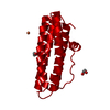
- Structure visualization
| Structure viewer | Molecule: MolmilJmol/JSmol |
|---|
- Downloads & links
-Download
| PDBx/mmCIF format | 2fjc.cif.gz | 466.2 KB | Display | PDBx/mmCIF format |
|---|---|---|---|---|
| PDB format | pdb2fjc.ent.gz | 386.6 KB | Display | PDB format |
| PDBx/mmJSON format | 2fjc.json.gz | Tree view | PDBx/mmJSON format | |
| Others |  Other downloads Other downloads |
-Validation report
| Arichive directory | https://data.pdbj.org/pub/pdb/validation_reports/fj/2fjcftp://data.pdbj.org/pub/pdb/validation_reports/fj/2fjc | HTTPS FTP |
|---|
-Related structure data
| Related structure data |  1ji4S S: Starting model for refinement |
|---|---|
| Similar structure data |


-Links
 PDBj
PDBj


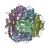
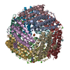
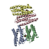
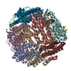
- Assembly
| Deposited unit | 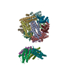
| ||||||||
|---|---|---|---|---|---|---|---|---|---|
| 1 | 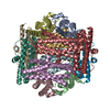
| ||||||||
| 2 | 
| ||||||||
| 3 | 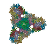
| ||||||||
| Unit cell |
|
-Components
| #1: Protein | Mass: 17252.652 Da / Num. of mol.: 16 Source method: isolated from a genetically manipulated source Source: (gene. exp.) Treponema pallidum (bacteria) / Gene: tpf1 / Plasmid: pSM214G-TpF1 / Production host: #2: Chemical | ChemComp-FE /   Mass: 55.845 Da / Num. of mol.: 16 / Source method: obtained synthetically / Formula: Fe Mass: 55.845 Da / Num. of mol.: 16 / Source method: obtained synthetically / Formula: Fe#3: Water | ChemComp-HOH / |  Mass: 18.015 Da / Num. of mol.: 517 / Source method: isolated from a natural source / Formula: H2O Mass: 18.015 Da / Num. of mol.: 517 / Source method: isolated from a natural source / Formula: H2O |
|---|
-Experimental details
-Experiment
| Experiment | Method: X-RAY DIFFRACTION / Number of used crystals: 1 |
|---|
- Sample preparation
| Crystal | Density Matthews: 2.77 Å3/Da / Density % sol: 55.58 % |
|---|---|
| Crystal grow | Temperature: 298 K / Method: vapor diffusion, sitting drop / pH: 7.5 Details: 10% PEG 6000, 8% ethylene glycol, 0.1 M Tris buffer, pH 7.5, VAPOR DIFFUSION, SITTING DROP, temperature 298K |
-Data collection
| Diffraction | Mean temperature: 100 K |
|---|---|
| Diffraction source | Source: SYNCHROTRON / Site: ELETTRA  / Beamline: 5.2R / Wavelength: 1.2 Å / Beamline: 5.2R / Wavelength: 1.2 Å |
| Detector | Type: MARRESEARCH / Detector: CCD / Date: Jun 1, 2004 |
| Radiation | Monochromator: Si 111 / Protocol: SINGLE WAVELENGTH / Monochromatic (M) / Laue (L): M / Scattering type: x-ray |
| Radiation wavelength | Wavelength: 1.2 Å / Relative weight: 1 |
| Reflection | Resolution: 2.45→185 Å / Num. all: 111030 / Num. obs: 111030 / % possible obs: 95.7 % / Observed criterion σ(F): 0 / Observed criterion σ(I): 0 / Redundancy: 5.1 % / Biso Wilson estimate: 56.6 Å2 / Rmerge(I) obs: 0.072 / Net I/σ(I): 8.5 |
| Reflection shell | Resolution: 2.45→2.55 Å / Redundancy: 3.9 % / Rmerge(I) obs: 0.37 / Mean I/σ(I) obs: 1.5 / Num. unique all: 11886 / % possible all: 70.8 |
- Processing
| Software |
| ||||||||||||||||||||||||||||||||||||||||||||||||||||||||||||||||||||||||||||||||
|---|---|---|---|---|---|---|---|---|---|---|---|---|---|---|---|---|---|---|---|---|---|---|---|---|---|---|---|---|---|---|---|---|---|---|---|---|---|---|---|---|---|---|---|---|---|---|---|---|---|---|---|---|---|---|---|---|---|---|---|---|---|---|---|---|---|---|---|---|---|---|---|---|---|---|---|---|---|---|---|---|---|
| Refinement | Method to determine structure: MOLECULAR REPLACEMENT Starting model: PDB entry 1JI4 Resolution: 2.5→59.37 Å / Rfactor Rfree error: 0.002 / Data cutoff high absF: 1010942.9 / Data cutoff low absF: 0 / Isotropic thermal model: RESTRAINED / Cross valid method: THROUGHOUT / σ(F): 0 / Stereochemistry target values: Engh & Huber
| ||||||||||||||||||||||||||||||||||||||||||||||||||||||||||||||||||||||||||||||||
| Solvent computation | Solvent model: FLAT MODEL / Bsol: 31.4013 Å2 / ksol: 0.349013 e/Å3 | ||||||||||||||||||||||||||||||||||||||||||||||||||||||||||||||||||||||||||||||||
| Displacement parameters | Biso mean: 40.4 Å2
| ||||||||||||||||||||||||||||||||||||||||||||||||||||||||||||||||||||||||||||||||
| Refine analyze |
| ||||||||||||||||||||||||||||||||||||||||||||||||||||||||||||||||||||||||||||||||
| Refinement step | Cycle: LAST / Resolution: 2.5→59.37 Å
| ||||||||||||||||||||||||||||||||||||||||||||||||||||||||||||||||||||||||||||||||
| Refine LS restraints |
| ||||||||||||||||||||||||||||||||||||||||||||||||||||||||||||||||||||||||||||||||
| Refine LS restraints NCS | NCS model details: CONSTR | ||||||||||||||||||||||||||||||||||||||||||||||||||||||||||||||||||||||||||||||||
| LS refinement shell | Resolution: 2.5→2.66 Å / Rfactor Rfree error: 0.008 / Total num. of bins used: 6
| ||||||||||||||||||||||||||||||||||||||||||||||||||||||||||||||||||||||||||||||||
| Xplor file |
|
