+ Open data
- Basic information
| Entry | Database: PDB / ID: 2fbe | ||||||
|---|---|---|---|---|---|---|---|
| Title | Crystal Structure of the PRYSPRY-domain | ||||||
 Components Components | PREDICTED: similar to ret finger protein-like 1 | ||||||
 Keywords Keywords | UNKNOWN FUNCTION / dimer / jellyroll beta-sandwich fold | ||||||
| Function / homology |  Function and homology information Function and homology informationubiquitin protein ligase activity / regulation of gene expression / innate immune response / zinc ion binding / nucleus / cytoplasm Similarity search - Function | ||||||
| Biological species |  Homo sapiens (human) Homo sapiens (human) | ||||||
| Method |  X-RAY DIFFRACTION / X-RAY DIFFRACTION /  SYNCHROTRON / SYNCHROTRON /  MAD / Resolution: 2.52 Å MAD / Resolution: 2.52 Å | ||||||
 Authors Authors | Gruetter, C. / Briand, C. / Capitani, G. / Mittl, P.R. / Gruetter, M.G. | ||||||
 Citation Citation |  Journal: Febs Lett. / Year: 2006 Journal: Febs Lett. / Year: 2006Title: Structure of the PRYSPRY-domain: Implications for autoinflammatory diseases Authors: Gruetter, C. / Briand, C. / Capitani, G. / Mittl, P.R. / Papin, S. / Tschopp, J. / Gruetter, M.G. | ||||||
| History |
|
- Structure visualization
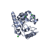
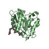
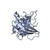
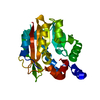
| Structure viewer | Molecule:  Molmil Molmil Jmol/JSmol Jmol/JSmol |
|---|
- Downloads & links
- Download
| PDBx/mmCIF format |  2fbe.cif.gz 2fbe.cif.gz | 166.2 KB | Display |  PDBx/mmCIF format PDBx/mmCIF format |
|---|---|---|---|---|
| PDB format |  pdb2fbe.ent.gz pdb2fbe.ent.gz | 133.8 KB | Display |  PDB format PDB format |
| PDBx/mmJSON format |  2fbe.json.gz 2fbe.json.gz | Tree view |  PDBx/mmJSON format PDBx/mmJSON format | |
| Others |  Other downloads Other downloads |
-Validation report
| Summary document |  2fbe_validation.pdf.gz 2fbe_validation.pdf.gz | 459 KB | Display |  wwPDB validaton report wwPDB validaton report |
|---|---|---|---|---|
| Full document |  2fbe_full_validation.pdf.gz 2fbe_full_validation.pdf.gz | 489.5 KB | Display | |
| Data in XML |  2fbe_validation.xml.gz 2fbe_validation.xml.gz | 39 KB | Display | |
| Data in CIF |  2fbe_validation.cif.gz 2fbe_validation.cif.gz | 50.9 KB | Display | |
| Arichive directory |  https://data.pdbj.org/pub/pdb/validation_reports/fb/2fbe https://data.pdbj.org/pub/pdb/validation_reports/fb/2fbe ftp://data.pdbj.org/pub/pdb/validation_reports/fb/2fbe ftp://data.pdbj.org/pub/pdb/validation_reports/fb/2fbe | HTTPS FTP |
-Related structure data
| Similar structure data |
|---|
- Links
Links
- Assembly
| Deposited unit | 
| ||||||||
|---|---|---|---|---|---|---|---|---|---|
| 1 | 
| ||||||||
| 2 | 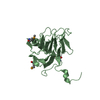
| ||||||||
| 3 | 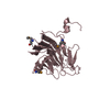
| ||||||||
| 4 | 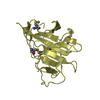
| ||||||||
| Unit cell |
| ||||||||
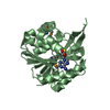
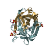
| Details | PRYSPRY can form a dimer at high concentration and shows a dimer in the crystal structure. The biological assembly could be a monomer or a dimer. |
- Components
| #1: Protein | Mass: 22431.736 Da / Num. of mol.: 4 / Fragment: PRYSPRY DOMAIN Source method: isolated from a genetically manipulated source Source: (gene. exp.)  Homo sapiens (human) / Gene: gene 19q13.4.1 / Plasmid: pGEX-6P1 / Species (production host): Escherichia coli / Production host: Homo sapiens (human) / Gene: gene 19q13.4.1 / Plasmid: pGEX-6P1 / Species (production host): Escherichia coli / Production host:  #2: Water | ChemComp-HOH / | Has protein modification | Y | |
|---|
-Experimental details
-Experiment
| Experiment | Method:  X-RAY DIFFRACTION / Number of used crystals: 2 X-RAY DIFFRACTION / Number of used crystals: 2 |
|---|
- Sample preparation
| Crystal | Density Matthews: 2.47 Å3/Da / Density % sol: 50.17 % |
|---|---|
| Crystal grow | Temperature: 277 K / Method: vapor diffusion, sitting drop / pH: 6.5 Details: 25% PEG 2000 MME, 0.55M KSCN, 0.1M cacodylate, pH 6.5, VAPOR DIFFUSION, SITTING DROP, temperature 277.0K |
-Data collection
| Diffraction |
| ||||||||||||||||||
|---|---|---|---|---|---|---|---|---|---|---|---|---|---|---|---|---|---|---|---|
| Diffraction source |
| ||||||||||||||||||
| Detector |
| ||||||||||||||||||
| Radiation |
| ||||||||||||||||||
| Radiation wavelength |
| ||||||||||||||||||
| Reflection | Resolution: 2.52→40 Å / Num. all: 31707 / Num. obs: 30566 / % possible obs: 96.4 % / Observed criterion σ(I): -3 / Redundancy: 4.3 % / Biso Wilson estimate: 52.1 Å2 / Rsym value: 0.081 / Net I/σ(I): 16.1 | ||||||||||||||||||
| Reflection shell | Resolution: 2.52→2.61 Å / Redundancy: 4.3 % / Mean I/σ(I) obs: 3.1 / Num. unique all: 3059 / Rsym value: 0.502 / % possible all: 99 |
- Processing
| Software |
| ||||||||||||||||||||||||||||||||||||
|---|---|---|---|---|---|---|---|---|---|---|---|---|---|---|---|---|---|---|---|---|---|---|---|---|---|---|---|---|---|---|---|---|---|---|---|---|---|
| Refinement | Method to determine structure:  MAD / Resolution: 2.52→30.46 Å / Rfactor Rfree error: 0.007 / Data cutoff high absF: 2409019.72 / Data cutoff low absF: 0 / Isotropic thermal model: RESTRAINED / Cross valid method: THROUGHOUT / σ(F): 0 / Stereochemistry target values: Engh & Huber MAD / Resolution: 2.52→30.46 Å / Rfactor Rfree error: 0.007 / Data cutoff high absF: 2409019.72 / Data cutoff low absF: 0 / Isotropic thermal model: RESTRAINED / Cross valid method: THROUGHOUT / σ(F): 0 / Stereochemistry target values: Engh & Huber
| ||||||||||||||||||||||||||||||||||||
| Solvent computation | Solvent model: FLAT MODEL / Bsol: 36.96 Å2 / ksol: 0.290646 e/Å3 | ||||||||||||||||||||||||||||||||||||
| Displacement parameters | Biso mean: 48.8 Å2
| ||||||||||||||||||||||||||||||||||||
| Refine analyze |
| ||||||||||||||||||||||||||||||||||||
| Refinement step | Cycle: LAST / Resolution: 2.52→30.46 Å
| ||||||||||||||||||||||||||||||||||||
| Refine LS restraints |
| ||||||||||||||||||||||||||||||||||||
| LS refinement shell | Resolution: 2.52→2.68 Å / Rfactor Rfree error: 0.024 / Total num. of bins used: 6
| ||||||||||||||||||||||||||||||||||||
| Xplor file |
|
 Movie
Movie Controller





 PDBj
PDBj


