 ムービー
ムービー コントローラー


+ データを開く

- 基本情報
基本情報
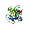
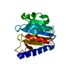
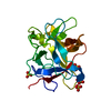
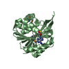
| 登録情報 | データベース: PDB / ID: 4wbc | |||||||||
|---|---|---|---|---|---|---|---|---|---|---|
| タイトル | 2.13 A STRUCTURE OF A KUNITZ-TYPE WINGED BEAN CHYMOTRYPSIN INHIBITOR PROTEIN | |||||||||
 要素 要素 | PROTEIN (CHYMOTRYPSIN INHIBITOR) | |||||||||
 キーワード キーワード | SERINE PROTEASE INHIBITOR | |||||||||
| 機能・相同性 |  機能・相同性情報 機能・相同性情報 | |||||||||
| 生物種 |   Psophocarpus tetragonolobus (マメ科) Psophocarpus tetragonolobus (マメ科) | |||||||||
| 手法 |  X線回折 / OTHER / 解像度: 2.138 Å X線回折 / OTHER / 解像度: 2.138 Å | |||||||||
 データ登録者 データ登録者 | Ravichandran, S. / Sen, U. / Chakrabarti, C. / Dattagupta, J.K. | |||||||||
 引用 引用 | ジャーナル: Acta Crystallogr.,Sect.D / 年: 1999 タイトル: Cryocrystallography of a Kunitz-type serine protease inhibitor: the 90 K structure of winged bean chymotrypsin inhibitor (WCI) at 2.13 A resolution. 著者: Ravichandran, S. / Sen, U. / Chakrabarti, C. / Dattagupta, J.K. #1: ジャーナル: Proteins / 年: 1999タイトル: Refined crystal structure (2.3 A) of a double-headed winged bean alpha-chymotrypsin inhibitor and location of its second reactive site. 著者: Dattagupta, J.K. / Podder, A. / Chakrabarti, C. / Sen, U. / Mukhopadhyay, D. / Dutta, S.K. / Singh, M. #2: ジャーナル: Acta Crystallogr.,Sect.D / 年: 1996タイトル: Structure of a Kunitz-type chymotrypsin from winged bean seeds at 2.95 A resolution. 著者: Dattagupta, J.K. / Podder, A. / Chakrabarti, C. / Sen, U. / Dutta, S.K. / Singh, M. #3: ジャーナル: J.Mol.Biol. / 年: 1990 タイトル: Crystallization and preliminary X-ray studies of psophocarpin B1, a chymotrypsin inhibitor from winged bean seeds. 著者: Dattagupta, J.K. / Chakrabarti, C. / Podder, A. / Dutta, S.K. / Singh, M. | |||||||||
| 履歴 |
|
- 構造の表示
構造の表示
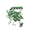
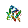
| 構造ビューア | 分子: MolmilJmol/JSmol |
|---|
- ダウンロードとリンク
-ダウンロード
| PDBx/mmCIF形式 | 4wbc.cif.gz | 53.2 KB | 表示 | PDBx/mmCIF形式 |
|---|---|---|---|---|
| PDB形式 | pdb4wbc.ent.gz | 38.9 KB | 表示 | PDB形式 |
| PDBx/mmJSON形式 | 4wbc.json.gz | ツリー表示 | PDBx/mmJSON形式 | |
| その他 |  その他のダウンロード その他のダウンロード |
-検証レポート
| アーカイブディレクトリ | https://data.pdbj.org/pub/pdb/validation_reports/wb/4wbcftp://data.pdbj.org/pub/pdb/validation_reports/wb/4wbc | HTTPS FTP |
|---|
-関連構造データ


-リンク
 PDBj
PDBj- 集合体
集合体
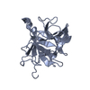
| 登録構造単位 | 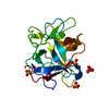
| ||||||||
|---|---|---|---|---|---|---|---|---|---|
| 1 |
| ||||||||
| 2 | 
| ||||||||
| 3 | 
| ||||||||
| 単位格子 |
| ||||||||
| Components on special symmetry positions |
|
-要素
| #1: タンパク質 | 分子量: 20266.850 Da / 分子数: 1 / 由来タイプ: 天然 / 由来: (天然) Psophocarpus tetragonolobus (マメ科) / 組織: SEED / 参照: UniProt: P10822 | ||||||||
|---|---|---|---|---|---|---|---|---|---|
| #2: 化合物 | ChemComp-SO4 /   分子量: 96.063 Da / 分子数: 5 / 由来タイプ: 合成 / 式: SO4 分子量: 96.063 Da / 分子数: 5 / 由来タイプ: 合成 / 式: SO4#3: 水 | ChemComp-HOH / |  分子量: 18.015 Da / 分子数: 176 / 由来タイプ: 天然 / 式: H2O 分子量: 18.015 Da / 分子数: 176 / 由来タイプ: 天然 / 式: H2OHas protein modification | Y | 非ポリマーの詳細 | THE PRESENCE OF SULPHATE IONS IN THE STRUCTURE IS DUE TO THE PRESENCE OF AMMONIUM SULPHATE SALT ...THE PRESENCE OF SULPHATE IONS IN THE STRUCTURE IS DUE TO THE PRESENCE OF AMMONIUM SULPHATE SALT USED IN THE CRYSTALLIZ | 配列の詳細 | REFERENCE: THE SEQUENCE OF WCI WAS TAKEN FROM SHIBATA, J. BIOCHEM. VOL. 134, 537-543, 1988. | |
-実験情報
-実験
| 実験 | 手法: X線回折 / 使用した結晶の数: 1 |
|---|
- 試料調製
試料調製
| 結晶 | マシュー密度: 2.69 Å3/Da / 溶媒含有率: 54 % 解説: THE CRYSTALS WERE MOUNTED IN A HAIR (HUMAN) LOOP AND FLASH FROZEN IN A STREAM OF LIQUID NITROGEN GAS STREAM AT 90 K FOR DATA COLLECTION. | ||||||||||||||||||||||||||||||||||||||||||||||||
|---|---|---|---|---|---|---|---|---|---|---|---|---|---|---|---|---|---|---|---|---|---|---|---|---|---|---|---|---|---|---|---|---|---|---|---|---|---|---|---|---|---|---|---|---|---|---|---|---|---|
| 結晶化 | pH: 5.4 詳細: PROTEIN WAS CRYSTALLIZED BY HANGING DROP VAPOUR DIFFUSION FROM 25% AMM. SULPHATE IN TRIS-HCL, 10MM NA ACETATE, 400 MM NACL AT PH 5.4 AGAINST 25% AMM. SULPHATE,60MM NA ACETATE AT 4 DEG. C. ...詳細: PROTEIN WAS CRYSTALLIZED BY HANGING DROP VAPOUR DIFFUSION FROM 25% AMM. SULPHATE IN TRIS-HCL, 10MM NA ACETATE, 400 MM NACL AT PH 5.4 AGAINST 25% AMM. SULPHATE,60MM NA ACETATE AT 4 DEG. C. CRYSTALS WERE THEN TRANSFERED TO 25% GLYCEROL (CRYOPROTECTANT) IN 25% AMM. SULPHATE, 10MM NA ACETATE BUFFER AT PH 5.4, BEFORE FLASH COOLING. | ||||||||||||||||||||||||||||||||||||||||||||||||
| 結晶 | *PLUS | ||||||||||||||||||||||||||||||||||||||||||||||||
| 結晶化 | *PLUS 温度: 4 ℃ / 手法: 蒸気拡散法, ハンギングドロップ法詳細: Dattagupta, J.K., (1999) Proteins: Struct., Funct., Genet., 35, 321 | ||||||||||||||||||||||||||||||||||||||||||||||||
| 溶液の組成 | *PLUS
|
-データ収集
| 回折 | 平均測定温度: 90 K |
|---|---|
| 放射光源 | 由来: 回転陽極 / タイプ: RIGAKU RU200 / 波長: 1.5418 |
| 検出器 | タイプ: MARRESEARCH / 検出器: IMAGE PLATE / 日付: 1997年5月1日 / 詳細: DOUBLE FOCUSSING MIRRORS |
| 放射 | モノクロメーター: NI FILTER / プロトコル: SINGLE WAVELENGTH / 単色(M)・ラウエ(L): M / 散乱光タイプ: x-ray |
| 放射波長 | 波長: 1.5418 Å / 相対比: 1 |
| 反射 | 解像度: 2.138→15 Å / Num. obs: 12963 / % possible obs: 96.3 % / Observed criterion σ(I): 0 / Biso Wilson estimate: 30.5 Å2 / Rmerge(I) obs: 0.069 / Net I/σ(I): 13 |
| 反射 シェル | 解像度: 2.13→2.18 Å / Rmerge(I) obs: 0.236 / % possible all: 64.5 |
| 反射 | *PLUS Num. measured all: 63933 |
- 解析
解析
| ソフトウェア |
| ||||||||||||||||||||||||||||||||||||||||||||||||||||||||||||||||||||||||||||||||||||
|---|---|---|---|---|---|---|---|---|---|---|---|---|---|---|---|---|---|---|---|---|---|---|---|---|---|---|---|---|---|---|---|---|---|---|---|---|---|---|---|---|---|---|---|---|---|---|---|---|---|---|---|---|---|---|---|---|---|---|---|---|---|---|---|---|---|---|---|---|---|---|---|---|---|---|---|---|---|---|---|---|---|---|---|---|---|
| 精密化 | 構造決定の手法: OTHER / 解像度: 2.138→9 Å / SU B: 3.02 / SU ML: 0.07 / 交差検証法: THROUGHOUT / σ(F): 0 / ESU R: 0.26 / ESU R Free: 0.22 詳細: PDB ENTRY 2WBC IS THE STARTING MODEL FOR REFINEMENT. INITIAL REFINEMENTS WERE DONE USING XPLOR 3.851
| ||||||||||||||||||||||||||||||||||||||||||||||||||||||||||||||||||||||||||||||||||||
| 原子変位パラメータ | Biso mean: 32.7 Å2 | ||||||||||||||||||||||||||||||||||||||||||||||||||||||||||||||||||||||||||||||||||||
| 精密化ステップ | サイクル: LAST / 解像度: 2.138→9 Å
| ||||||||||||||||||||||||||||||||||||||||||||||||||||||||||||||||||||||||||||||||||||
| 拘束条件 |
| ||||||||||||||||||||||||||||||||||||||||||||||||||||||||||||||||||||||||||||||||||||
| ソフトウェア | *PLUS 名称: REFMAC / 分類: refinement | ||||||||||||||||||||||||||||||||||||||||||||||||||||||||||||||||||||||||||||||||||||
| 精密化 | *PLUS 最低解像度: 9 Å / σ(F): 0 / % reflection Rfree: 10 % / Rfactor obs: 0.2 | ||||||||||||||||||||||||||||||||||||||||||||||||||||||||||||||||||||||||||||||||||||
| 溶媒の処理 | *PLUS | ||||||||||||||||||||||||||||||||||||||||||||||||||||||||||||||||||||||||||||||||||||
| 原子変位パラメータ | *PLUS Biso mean: 32.7 Å2 | ||||||||||||||||||||||||||||||||||||||||||||||||||||||||||||||||||||||||||||||||||||
| 拘束条件 | *PLUS
|
