 Movie
Movie Controller

[English] 日本語
 Yorodumi
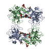
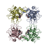
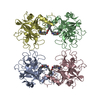
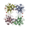
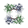
- PDB-2f9p: Crystal Structure of the Recombinant Human Alpha I Tryptase Mutan... -
+ Open data

- Basic information
| Entry | Database: PDB / ID: 2f9p | |||||||||
|---|---|---|---|---|---|---|---|---|---|---|
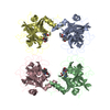
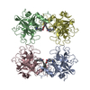
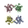
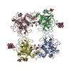
| Title | Crystal Structure of the Recombinant Human Alpha I Tryptase Mutant D216G in Complex with Leupeptin | |||||||||
 Components Components |
| |||||||||
 Keywords Keywords | HYDROLASE/HYDROLASE INHIBITOR / serine proteinase / leupeptin / trypsin-like / difucosylation / HYDROLASE / HYDROLASE-HYDROLASE INHIBITOR COMPLEX | |||||||||
| Function / homology |  Function and homology information Function and homology informationtryptase / Activation of Matrix Metalloproteinases / extracellular matrix disassembly / serine-type peptidase activity / defense response / serine-type endopeptidase activity / proteolysis / : / extracellular region / identical protein binding Similarity search - Function | |||||||||
| Biological species |  Homo sapiens (human) Homo sapiens (human) Actinomycetes Streptomyces roseus MA 839-A1 (bacteria) Actinomycetes Streptomyces roseus MA 839-A1 (bacteria) | |||||||||
| Method |  X-RAY DIFFRACTION / MOLECULAR REPLACEMENT / Resolution: 2.3 Å X-RAY DIFFRACTION / MOLECULAR REPLACEMENT / Resolution: 2.3 Å | |||||||||
 Authors Authors | Rohr, K.B. / Selwood, T. / Marquardt, U. / Huber, R. / Schechter, N.M. / Bode, W. / Than, M.E. | |||||||||
 Citation Citation | Journal: J.Mol.Biol. / Year: 2005 Title: X-ray Structures of Free and Leupeptin-complexed Human alpha I-Tryptase Mutants: Indication for an alpha to beta-Tryptase Transition Authors: Rohr, K.B. / Selwood, T. / Marquardt, U. / Huber, R. / Schechter, N.M. / Bode, W. / Than, M.E. | |||||||||
| History |
|
- Structure visualization
| Structure viewer | Molecule: MolmilJmol/JSmol |
|---|
- Downloads & links
-Download
| PDBx/mmCIF format | 2f9p.cif.gz | 221 KB | Display | PDBx/mmCIF format |
|---|---|---|---|---|
| PDB format | pdb2f9p.ent.gz | 176.5 KB | Display | PDB format |
| PDBx/mmJSON format | 2f9p.json.gz | Tree view | PDBx/mmJSON format | |
| Others |  Other downloads Other downloads |
-Validation report
| Arichive directory | https://data.pdbj.org/pub/pdb/validation_reports/f9/2f9pftp://data.pdbj.org/pub/pdb/validation_reports/f9/2f9p | HTTPS FTP |
|---|
-Related structure data
| Related structure data |  2f9nSC  2f9oC S: Starting model for refinement C: citing same article ( |
|---|---|
| Similar structure data |

-Links
 PDBj
PDBj
- Assembly
| Deposited unit | 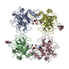
| ||||||||
|---|---|---|---|---|---|---|---|---|---|
| 1 |
| ||||||||
| Unit cell |
|
-Components
-Protein / Protein/peptide , 2 types, 8 molecules ABCDEFGH
| #1: Protein | Mass: 27660.691 Da / Num. of mol.: 4 / Fragment: residues 31-275 / Mutation: D216G Source method: isolated from a genetically manipulated source Source: (gene. exp.) Homo sapiens (human) / Gene: TPSAB1, TPS1 / Plasmid: pAcGP67B / Production host:   Spodoptera frugiperda (fall armyworm) / Strain (production host): Hi5 Spodoptera frugiperda (fall armyworm) / Strain (production host): Hi5References: GenBank: 1182067, UniProt: Q15661*PLUS, tryptase #2: Protein/peptide | 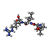  Type: Oligopeptide / Class: Enzyme inhibitor / Mass: 429.578 Da / Num. of mol.: 4 / Source method: obtained synthetically Type: Oligopeptide / Class: Enzyme inhibitor / Mass: 429.578 Da / Num. of mol.: 4 / Source method: obtained syntheticallySource: (synth.) Actinomycetes Streptomyces roseus MA 839-A1 (bacteria)References: NOR: NOR00487, LEUPEPTIN |
|---|
-Sugars , 2 types, 2 molecules 
| #3: Polysaccharide | alpha-L-fucopyranose-(1-3)-2-acetamido-2-deoxy-beta-D-glucopyranose Source method: isolated from a genetically manipulated source |
|---|---|
| #5: Sugar | ChemComp-NAG /  Type: D-saccharide, beta linking / Mass: 221.208 Da / Num. of mol.: 1 Type: D-saccharide, beta linking / Mass: 221.208 Da / Num. of mol.: 1Source method: isolated from a genetically manipulated source Formula: C8H15NO6 |
-Non-polymers , 2 types, 506 molecules 

| #4: Chemical | ChemComp-BU3 / (  Mass: 90.121 Da / Num. of mol.: 4 / Source method: obtained synthetically / Formula: C4H10O2 Mass: 90.121 Da / Num. of mol.: 4 / Source method: obtained synthetically / Formula: C4H10O2#6: Water | ChemComp-HOH / | Mass: 18.015 Da / Num. of mol.: 502 / Source method: isolated from a natural source / Formula: H2O |
|---|
-Details
| Compound details | THE LEUPEPTIN IS COVALENTLY| Has protein modification | Y | Sequence details | THERE IS A DISCREPANCY IN THE NORINE AND PDB NUMBERING, AS NORINE COUNTS ACE AND LEU TOGETHER AS ...THERE IS A DISCREPANC | |
|---|
-Experimental details
-Experiment
| Experiment | Method: X-RAY DIFFRACTION / Number of used crystals: 1 |
|---|
- Sample preparation
| Crystal | Density Matthews: 2.66 Å3/Da / Density % sol: 53.78 % |
|---|---|
| Crystal grow | Temperature: 293 K / Method: vapor diffusion, sitting drop / pH: 6.8 Details: 28% PEG1500, pH 6.80, VAPOR DIFFUSION, SITTING DROP, temperature 293K |
-Data collection
| Diffraction | Mean temperature: 100 K |
|---|---|
| Diffraction source | Source: ROTATING ANODE / Type: RIGAKU / Wavelength: 1.54 |
| Detector | Type: MARRESEARCH / Detector: IMAGE PLATE / Date: Nov 1, 2004 |
| Radiation | Protocol: SINGLE WAVELENGTH / Monochromatic (M) / Laue (L): M / Scattering type: x-ray |
| Radiation wavelength | Wavelength: 1.54 Å / Relative weight: 1 |
| Reflection | Resolution: 2.3→16 Å / Num. obs: 53746 / % possible obs: 99.6 % / Biso Wilson estimate: 25.6 Å2 / Rmerge(I) obs: 0.091 / Net I/σ(I): 15.1 |
| Reflection shell | Resolution: 2.3→2.38 Å / Rmerge(I) obs: 0.36 / Mean I/σ(I) obs: 3.3 / % possible all: 98.3 |
- Processing
| Software |
| ||||||||||||||||||||||||||||||||||||||||||||||||||||||||||||
|---|---|---|---|---|---|---|---|---|---|---|---|---|---|---|---|---|---|---|---|---|---|---|---|---|---|---|---|---|---|---|---|---|---|---|---|---|---|---|---|---|---|---|---|---|---|---|---|---|---|---|---|---|---|---|---|---|---|---|---|---|---|
| Refinement | Method to determine structure: MOLECULAR REPLACEMENT Starting model: PDB ENTRY 2F9N Resolution: 2.3→15 Å / Rfactor Rfree error: 0.005 / Isotropic thermal model: ANISOTROPIC / Cross valid method: THROUGHOUT / σ(F): 0 / Stereochemistry target values: ENGH & HUBER
| ||||||||||||||||||||||||||||||||||||||||||||||||||||||||||||
| Solvent computation | Solvent model: FLAT MODEL / Bsol: 35.23 Å2 / ksol: 0.34 e/Å3 | ||||||||||||||||||||||||||||||||||||||||||||||||||||||||||||
| Displacement parameters | Biso mean: 27.1 Å2
| ||||||||||||||||||||||||||||||||||||||||||||||||||||||||||||
| Refine analyze |
| ||||||||||||||||||||||||||||||||||||||||||||||||||||||||||||
| Refinement step | Cycle: LAST / Resolution: 2.3→15 Å
| ||||||||||||||||||||||||||||||||||||||||||||||||||||||||||||
| Refine LS restraints |
| ||||||||||||||||||||||||||||||||||||||||||||||||||||||||||||
| LS refinement shell | Resolution: 2.3→2.4 Å /
|
