 Movie
Movie Controller

[English] 日本語
 Yorodumi
- PDB-2f6i: Crystal structure of the ClpP protease catalytic domain from Plas... -
+ Open data

- Basic information
| Entry | Database: PDB / ID: 2f6i | ||||||
|---|---|---|---|---|---|---|---|
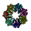
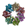
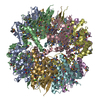
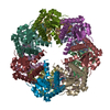
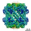
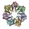
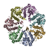
| Title | Crystal structure of the ClpP protease catalytic domain from Plasmodium falciparum | ||||||
 Components Components | ATP-dependent CLP protease, putative | ||||||
 Keywords Keywords | HYDROLASE / Clp protease / structural genomics / Structural Genomics Consortium / SGC | ||||||
| Function / homology |  Function and homology information Function and homology informationapicoplast / endopeptidase Clp complex / ATP-dependent peptidase activity / protein quality control for misfolded or incompletely synthesized proteins / ATPase binding / serine-type endopeptidase activity / hydrolase activity / proteolysis Similarity search - Function | ||||||
| Biological species |  | ||||||
| Method |  X-RAY DIFFRACTION / SYNCHROTRON / MOLECULAR REPLACEMENT / Resolution: 2.45 Å X-RAY DIFFRACTION / SYNCHROTRON / MOLECULAR REPLACEMENT / Resolution: 2.45 Å | ||||||
 Authors Authors | Mulichak, A. / Loppnau, P. / Bray, J. / Amani, M. / Vedadi, M. / Wasney, G. / Finerty, P. / Sundstrom, M. / Weigelt, J. / Edwards, A. ...Mulichak, A. / Loppnau, P. / Bray, J. / Amani, M. / Vedadi, M. / Wasney, G. / Finerty, P. / Sundstrom, M. / Weigelt, J. / Edwards, A. / Arrowsmith, C. / Hui, R. / Plotnikova, O. / Structural Genomics Consortium (SGC) | ||||||
 Citation Citation | Journal: J.Mol.Biol. / Year: 2010 Title: The Clp chaperones and proteases of the human malaria parasite Plasmodium falciparum. Authors: El Bakkouri, M. / Pow, A. / Mulichak, A. / Cheung, K.L. / Artz, J.D. / Amani, M. / Fell, S. / de Koning-Ward, T.F. / Goodman, C.D. / McFadden, G.I. / Ortega, J. / Hui, R. / Houry, W.A. #1: Journal: Mol.Biochem.Parasitol. / Year: 2007Title: Genome-scale protein expression and structural biology of Plasmodium falciparum and related Apicomplexan organisms. Authors: Vedadi, M. / Lew, J. / Artz, J. / Amani, M. / Zhao, Y. / Dong, A. / Wasney, G.A. / Gao, M. / Hills, T. / Brokx, S. / Qiu, W. / Sharma, S. / Diassiti, A. / Alam, Z. / Melone, M. / Mulichak, A. ...Authors: Vedadi, M. / Lew, J. / Artz, J. / Amani, M. / Zhao, Y. / Dong, A. / Wasney, G.A. / Gao, M. / Hills, T. / Brokx, S. / Qiu, W. / Sharma, S. / Diassiti, A. / Alam, Z. / Melone, M. / Mulichak, A. / Wernimont, A. / Bray, J. / Loppnau, P. / Plotnikova, O. / Newberry, K. / Sundararajan, E. / Houston, S. / Walker, J. / Tempel, W. / Bochkarev, A. / Kozieradzki, I. / Edwards, A. / Arrowsmith, C. / Roos, D. / Kain, K. / Hui, R. | ||||||
| History |
|
- Structure visualization
| Structure viewer | Molecule: MolmilJmol/JSmol |
|---|
- Downloads & links
-Download
| PDBx/mmCIF format | 2f6i.cif.gz | 258.1 KB | Display | PDBx/mmCIF format |
|---|---|---|---|---|
| PDB format | pdb2f6i.ent.gz | 207.3 KB | Display | PDB format |
| PDBx/mmJSON format | 2f6i.json.gz | Tree view | PDBx/mmJSON format | |
| Others |  Other downloads Other downloads |
-Validation report
| Arichive directory | https://data.pdbj.org/pub/pdb/validation_reports/f6/2f6iftp://data.pdbj.org/pub/pdb/validation_reports/f6/2f6i | HTTPS FTP |
|---|
-Related structure data
| Related structure data | 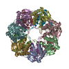 1tyfS S: Starting model for refinement |
|---|---|
| Similar structure data |

-Links
 PDBj
PDBj- Assembly
| Deposited unit | 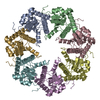
| ||||||||
|---|---|---|---|---|---|---|---|---|---|
| 1 | 
| ||||||||
| Unit cell |
| ||||||||
| Details | The biological assembly is a tetradecamer generated from the heptamer in the asymmetric unit by the operation: -X, Y, -Z+1/2 |
-Components
| #1: Protein | Mass: 24870.557 Da / Num. of mol.: 7 Source method: isolated from a genetically manipulated source Source: (gene. exp.) Plasmid: pET-28a vector customized for thrombin cleavage and LIC Production host:  #2: Water | ChemComp-HOH / |  Mass: 18.015 Da / Num. of mol.: 248 / Source method: isolated from a natural source / Formula: H2O Mass: 18.015 Da / Num. of mol.: 248 / Source method: isolated from a natural source / Formula: H2O |
|---|
-Experimental details
-Experiment
| Experiment | Method: X-RAY DIFFRACTION / Number of used crystals: 1 |
|---|
- Sample preparation
| Crystal | Density Matthews: 3.11 Å3/Da / Density % sol: 60.43 % |
|---|---|
| Crystal grow | Temperature: 298 K / Method: vapor diffusion, hanging drop / pH: 7 Details: PEG MME 550, Ammonium sulfate, cacodylate buffer., pH 7.0, VAPOR DIFFUSION, HANGING DROP, temperature 298K |
-Data collection
| Diffraction | Mean temperature: 100 K |
|---|---|
| Diffraction source | Source: SYNCHROTRON / Site: NSLS  / Beamline: X25 / Wavelength: 1.1 Å / Beamline: X25 / Wavelength: 1.1 Å |
| Detector | Type: ADSC QUANTUM 315 / Detector: CCD / Date: Sep 17, 2005 |
| Radiation | Protocol: SINGLE WAVELENGTH / Monochromatic (M) / Laue (L): M / Scattering type: x-ray |
| Radiation wavelength | Wavelength: 1.1 Å / Relative weight: 1 |
| Reflection | Resolution: 2.45→50 Å / Num. all: 78895 / Num. obs: 78895 / % possible obs: 99.2 % / Observed criterion σ(F): 0 / Observed criterion σ(I): 0 / Redundancy: 7.5 % / Rsym value: 0.078 |
| Reflection shell | Resolution: 2.45→2.54 Å / Redundancy: 4.5 % / Num. unique all: 7777 / Rsym value: 0.316 / % possible all: 98.5 |
- Processing
| Software |
| ||||||||||||||||||||
|---|---|---|---|---|---|---|---|---|---|---|---|---|---|---|---|---|---|---|---|---|---|
| Refinement | Method to determine structure: MOLECULAR REPLACEMENT Starting model: PDB entry 1TYF Resolution: 2.45→30 Å / Cross valid method: THROUGHOUT / σ(F): 0 / Stereochemistry target values: Engh & Huber Details: The following loops are unobserved and omitted from the model: Chain A 297-304, Chain B 290-304, Chain C 298-303, Chain D 297-303, Chain E 297-304, Chain F 292-303. Side chain atoms of ...Details: The following loops are unobserved and omitted from the model: Chain A 297-304, Chain B 290-304, Chain C 298-303, Chain D 297-303, Chain E 297-304, Chain F 292-303. Side chain atoms of residues listed in Remark 470 are unobserved in electron density maps and are omitted from model.
| ||||||||||||||||||||
| Refine analyze | Luzzati coordinate error obs: 0.34 Å / Luzzati d res low obs: 5 Å / Luzzati sigma a obs: 0.39 Å | ||||||||||||||||||||
| Refinement step | Cycle: LAST / Resolution: 2.45→30 Å
| ||||||||||||||||||||
| Refine LS restraints |
|
