 Movie
Movie Controller

[English] 日本語
 Yorodumi
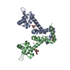
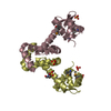
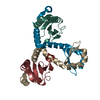
- PDB-2f5f: Bacillus subtilis manganese transport regulator (MNTR) bound to m... -
+ Open data

- Basic information
| Entry | Database: PDB / ID: 2f5f | ||||||
|---|---|---|---|---|---|---|---|
| Title | Bacillus subtilis manganese transport regulator (MNTR) bound to manganese, AC conformation, pH 8.5 | ||||||
 Components Components | Transcriptional regulator mntR | ||||||
 Keywords Keywords | TRANSCRIPTION / HELIX-TURN-HELIX / DNA-BINDING PROTEIN / METALLOREGULATORY PROTEIN | ||||||
| Function / homology |  Function and homology information Function and homology informationintracellular manganese ion homeostasis / manganese ion binding / protein dimerization activity / DNA-binding transcription factor activity / DNA-templated transcription / DNA binding / cytoplasm Similarity search - Function | ||||||
| Biological species |  | ||||||
| Method |  X-RAY DIFFRACTION / MOLECULAR REPLACEMENT / Resolution: 2.4 Å X-RAY DIFFRACTION / MOLECULAR REPLACEMENT / Resolution: 2.4 Å | ||||||
 Authors Authors | Kliegman, J.I. / Griner, S.L. / Helmann, J.D. / Brennan, R.G. / Glasfeld, A. | ||||||
 Citation Citation | Journal: Biochemistry / Year: 2006 Title: Structural Basis for the Metal-Selective Activation of the Manganese Transport Regulator of Bacillus subtilis. Authors: Kliegman, J.I. / Griner, S.L. / Helmann, J.D. / Brennan, R.G. / Glasfeld, A. | ||||||
| History |
| ||||||
| Remark 999 | SEQUENCE THE AUTHOR MAINTAINS THAT THE CORRECT RESIDUE AT THIS LOCATION SHOULD BE GLU. |
- Structure visualization
| Structure viewer | Molecule: MolmilJmol/JSmol |
|---|
- Downloads & links
-Download
| PDBx/mmCIF format | 2f5f.cif.gz | 68.5 KB | Display | PDBx/mmCIF format |
|---|---|---|---|---|
| PDB format | pdb2f5f.ent.gz | 50.9 KB | Display | PDB format |
| PDBx/mmJSON format | 2f5f.json.gz | Tree view | PDBx/mmJSON format | |
| Others |  Other downloads Other downloads |
-Validation report
| Arichive directory | https://data.pdbj.org/pub/pdb/validation_reports/f5/2f5fftp://data.pdbj.org/pub/pdb/validation_reports/f5/2f5f | HTTPS FTP |
|---|
-Related structure data
| Related structure data | 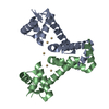 2ev0C 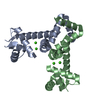 2ev5C  2ev6C 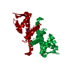 2f5cC 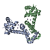 2f5dC 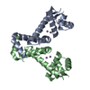 2f5eC 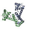 1on1S S: Starting model for refinement C: citing same article ( |
|---|---|
| Similar structure data |


-Links
 PDBj
PDBj- Assembly
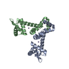
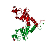
| Deposited unit | 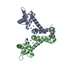
| ||||||||
|---|---|---|---|---|---|---|---|---|---|
| 1 |
| ||||||||
| Unit cell |
|
-Components
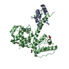
| #1: Protein | Mass: 16787.133 Da / Num. of mol.: 2 Source method: isolated from a genetically manipulated source Source: (gene. exp.) #2: Chemical | ChemComp-MN /   Mass: 54.938 Da / Num. of mol.: 4 / Source method: obtained synthetically / Formula: Mn Mass: 54.938 Da / Num. of mol.: 4 / Source method: obtained synthetically / Formula: Mn#3: Water | ChemComp-HOH / |  Mass: 18.015 Da / Num. of mol.: 37 / Source method: isolated from a natural source / Formula: H2O Mass: 18.015 Da / Num. of mol.: 37 / Source method: isolated from a natural source / Formula: H2O |
|---|
-Experimental details
-Experiment
| Experiment | Method: X-RAY DIFFRACTION / Number of used crystals: 1 |
|---|
- Sample preparation
| Crystal | Density Matthews: 2.63 Å3/Da / Density % sol: 53.27 % |
|---|---|
| Crystal grow | Temperature: 293 K / Method: vapor diffusion, hanging drop / pH: 8.5 Details: 30% PEG 400, 0.15 M LITHIUM SULFATE, PH 8.5, VAPOR DIFFUSION, HANGING DROP, TEMPERATURE 293K, pH 8.50 |
-Data collection
| Diffraction | Mean temperature: 295 K |
|---|---|
| Diffraction source | Source: ROTATING ANODE / Type: RIGAKU RU300 / Wavelength: 1.5418 |
| Detector | Type: RIGAKU RAXIS IV / Detector: IMAGE PLATE / Date: Jun 17, 2003 |
| Radiation | Monochromator: OSMIC MIRRORS / Protocol: SINGLE WAVELENGTH / Monochromatic (M) / Laue (L): M / Scattering type: x-ray |
| Radiation wavelength | Wavelength: 1.5418 Å / Relative weight: 1 |
| Reflection | Resolution: 2.4→31.7 Å / Num. obs: 13217 / % possible obs: 95 % / Observed criterion σ(I): 0 / Biso Wilson estimate: 24.1 Å2 / Rmerge(I) obs: 0.087 / Net I/σ(I): 5.2 |
| Reflection shell | Resolution: 2.4→2.55 Å / Rmerge(I) obs: 0.342 / Mean I/σ(I) obs: 2 / % possible all: 93.8 |
- Processing
| Software |
| ||||||||||||||||||||||||||||||||||||||||||||||||||||||||||||
|---|---|---|---|---|---|---|---|---|---|---|---|---|---|---|---|---|---|---|---|---|---|---|---|---|---|---|---|---|---|---|---|---|---|---|---|---|---|---|---|---|---|---|---|---|---|---|---|---|---|---|---|---|---|---|---|---|---|---|---|---|---|
| Refinement | Method to determine structure: MOLECULAR REPLACEMENT Starting model: PDB ENTRY 1ON1 Resolution: 2.4→31.7 Å / Rfactor Rfree error: 0.01 / Data cutoff high absF: 895970.2 / Data cutoff high rms absF: 895970.2 / Data cutoff low absF: 0 / Isotropic thermal model: RESTRAINED / Cross valid method: THROUGHOUT / σ(F): 0
| ||||||||||||||||||||||||||||||||||||||||||||||||||||||||||||
| Solvent computation | Solvent model: FLAT MODEL / Bsol: 43.8287 Å2 / ksol: 0.326559 e/Å3 | ||||||||||||||||||||||||||||||||||||||||||||||||||||||||||||
| Displacement parameters | Biso mean: 43.7 Å2
| ||||||||||||||||||||||||||||||||||||||||||||||||||||||||||||
| Refine analyze |
| ||||||||||||||||||||||||||||||||||||||||||||||||||||||||||||
| Refinement step | Cycle: LAST / Resolution: 2.4→31.7 Å
| ||||||||||||||||||||||||||||||||||||||||||||||||||||||||||||
| Refine LS restraints |
| ||||||||||||||||||||||||||||||||||||||||||||||||||||||||||||
| LS refinement shell | Resolution: 2.4→2.55 Å / Rfactor Rfree error: 0.033 / Total num. of bins used: 6
| ||||||||||||||||||||||||||||||||||||||||||||||||||||||||||||
| Xplor file |
|
