[English] 日本語
Yorodumi
- PDB-1q1k: Structure of ATP-phosphoribosyltransferase from E. coli complexed... -

+
Open data


ID or keywords:

Loading...

-
Basic information

Entry
Database: PDB / ID: 1q1k
TitleStructure of ATP-phosphoribosyltransferase from E. coli complexed with PR-ATP
ComponentsATP phosphoribosyltransferase
KeywordsTRANSFERASE / histidine biosynthesis / PRPP binding / PR-ATP inhibition
Function / homology
Function and homology information


ATP phosphoribosyltransferase / ATP phosphoribosyltransferase activity / histidine biosynthetic process / magnesium ion binding / ATP binding / cytosol
Similarity search - Function
ATP phosphoribosyltransferase HisG, long form / Histidine biosynthesis HisG, C-terminal / HisG, C-terminal domain / ATP phosphoribosyltransferase HisG / ATP phosphoribosyltransferase, catalytic domain / ATP phosphoribosyltransferase, conserved site / ATP phosphoribosyltransferase / ATP phosphoribosyltransferase signature. / Alpha-Beta Plaits - #120 / Nitrogen regulatory PII-like, alpha/beta ...ATP phosphoribosyltransferase HisG, long form / Histidine biosynthesis HisG, C-terminal / HisG, C-terminal domain / ATP phosphoribosyltransferase HisG / ATP phosphoribosyltransferase, catalytic domain / ATP phosphoribosyltransferase, conserved site / ATP phosphoribosyltransferase / ATP phosphoribosyltransferase signature. / Alpha-Beta Plaits - #120 / Nitrogen regulatory PII-like, alpha/beta / Nitrogen regulatory protein PII/ATP phosphoribosyltransferase, C-terminal / Periplasmic binding protein-like II / D-Maltodextrin-Binding Protein; domain 2 / Alpha-Beta Plaits / 2-Layer Sandwich / 3-Layer(aba) Sandwich / Alpha Beta
Similarity search - Domain/homology
PHOSPHORIBOSYL ATP / L(+)-TARTARIC ACID / ATP phosphoribosyltransferase
Similarity search - Component
Biological speciesEscherichia coli (E. coli)
MethodX-RAY DIFFRACTION / SYNCHROTRON / MOLECULAR REPLACEMENT / Resolution: 2.9 Å
AuthorsLohkamp, B. / McDermott, G. / Coggins, J.R. / Lapthorn, A.J.
CitationJournal: J.Mol.Biol. / Year: 2004
Title: The structure of Escherichia coli ATP-phosphoribosyltransferase: identification of substrate binding sites and mode of AMP inhibition
Authors: Lohkamp, B. / McDermott, G. / Campbell, S.A. / Coggins, J.R. / Lapthorn, A.J.
History
DepositionJul 21, 2003Deposition site: RCSB / Processing site: RCSB
Revision 1.0Mar 2, 2004Provider: repository / Type: Initial release
Revision 1.1Apr 29, 2008Group: Version format compliance
Revision 1.2Jul 13, 2011Group: Derived calculations / Version format compliance
Revision 1.3May 3, 2017Group: Non-polymer description
Revision 1.4Aug 16, 2023Group: Advisory / Data collection ...Advisory / Data collection / Database references / Derived calculations / Refinement description
Category: chem_comp_atom / chem_comp_bond ...chem_comp_atom / chem_comp_bond / database_2 / pdbx_initial_refinement_model / pdbx_unobs_or_zero_occ_atoms / struct_conn / struct_site
Item: _database_2.pdbx_DOI / _database_2.pdbx_database_accession ..._database_2.pdbx_DOI / _database_2.pdbx_database_accession / _struct_conn.pdbx_leaving_atom_flag / _struct_site.pdbx_auth_asym_id / _struct_site.pdbx_auth_comp_id / _struct_site.pdbx_auth_seq_id
Remark 600
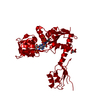
HETEROGEN The ligand PRT is a fragment of Phosphoribosyl ATP with the ribose and triphosphate ...HETEROGEN The ligand PRT is a fragment of Phosphoribosyl ATP with the ribose and triphosphate either disordered or cleaved within the crystal. The authors modelled the ribose and triphosphate with zero occupancy.

-
Structure visualization

Structure viewerMolecule:
MolmilJmol/JSmol

Downloads & links

-
Assembly

Deposited unit
A: ATP phosphoribosyltransferase
hetero molecules


Theoretical massNumber of molelcules
Total (without water)34,4284
Polymers33,4091
Non-polymers1,0193
Water0
1
A: ATP phosphoribosyltransferase
hetero molecules
x 6


Theoretical massNumber of molelcules
Total (without water)206,56924
Polymers200,4526
Non-polymers6,11718
Water0
TypeNameSymmetry operationNumber
identity operation1_555x,y,z1
crystal symmetry operation2_655-y+1,x-y,z1
crystal symmetry operation3_665-x+y+1,-x+1,z1
crystal symmetry operation16_545y+1/3,x-1/3,-z+2/31
crystal symmetry operation17_555x-y+1/3,-y+2/3,-z+2/31
crystal symmetry operation18_655-x+4/3,-x+y+2/3,-z+2/31
Buried area28510 Å2
ΔGint-165 kcal/mol
Surface area72260 Å2
MethodPISA, PQS
Unit cell
Length a, b, c (Å)132.100, 132.100, 113.379
Angle α, β, γ (deg.)90.00, 90.00, 120.00
Int Tables number155
Space group name H-MH32
Details
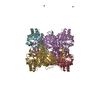
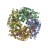
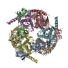
The biological assembly is a hexamer generated from the monomer in the asymmetric unit by the operations: -y, x-y, z; -x+y, -x, z; 1/3+y, 2/3+x, 2/3-z; 1/3+x-y, 2/3-y, 2/3-z and 1/3-x, 2/3-x+y, 2/3-z.

-
Components

#1: Protein ATP phosphoribosyltransferase /


Mass: 33408.746 Da / Num. of mol.: 1
Source method: isolated from a genetically manipulated source
Source: (gene. exp.) Escherichia coli (E. coli) / Gene: HISG / Plasmid: pTB361 / Production host: Escherichia coli (E. coli) / Strain (production host): B824(DE3)pLysS / References: UniProt: P60757, ATP phosphoribosyltransferase
#2: Chemical ChemComp-PRT / PHOSPHORIBOSYL ATP / [[(2~{R},3~{S},4~{R},5~{R})-5-[6-azanylidene-1-[(2~{R},3~{R},4~{S},5~{R})-3,4-bis(oxidanyl)-5-(phosphonooxymethyl)oxolan-2-yl]purin-9-yl]-3,4-bis(oxidanyl)oxolan-2-yl]methoxy-oxidanyl-phosphoryl] phosphono hydrogen phosphate


Mass: 719.276 Da / Num. of mol.: 1 / Source method: obtained synthetically / Formula: C15H25N5O20P4
#3: Chemical ChemComp-TLA / L(+)-TARTARIC ACID / Tartaric acid


Mass: 150.087 Da / Num. of mol.: 2 / Source method: obtained synthetically / Formula: C4H6O6

-
Experimental details

-
Experiment

ExperimentMethod: X-RAY DIFFRACTION / Number of used crystals: 1

-
Sample preparation

CrystalDensity Matthews: 2.85 Å3/Da / Density % sol: 56.82 %
Crystal growTemperature: 298 K / Method: vapor diffusion, sitting drop / pH: 5.6
Details: sodium tartrate, magnesium chloride, sodium citrate, pH 5.6, VAPOR DIFFUSION, SITTING DROP, temperature 298K

-
Data collection

DiffractionMean temperature: 100 K
Diffraction sourceSource: SYNCHROTRON / Site: SRS / Beamline: PX9.6 / Wavelength: 0.86 Å
DetectorType: ADSC QUANTUM 4 / Detector: CCD / Date: Jul 5, 2002
RadiationMonochromator: Si(111) / Protocol: SINGLE WAVELENGTH / Monochromatic (M) / Laue (L): M / Scattering type: x-ray
Radiation wavelengthWavelength: 0.86 Å / Relative weight: 1
ReflectionResolution: 2.9→50 Å / Num. all: 8521 / Num. obs: 8521 / % possible obs: 99.3 % / Observed criterion σ(F): 2 / Observed criterion σ(I): 2 / Redundancy: 4.2 % / Rmerge(I) obs: 0.058 / Net I/σ(I): 16.9
Reflection shellResolution: 2.9→3.02 Å / Redundancy: 4.2 % / Rmerge(I) obs: 0.581 / Mean I/σ(I) obs: 2 / % possible all: 99.9

-
Processing

Software
NameVersionClassification
REFMAC5.1.24refinement
DENZOdata reduction
SCALEPACKdata scaling
AMoREphasing
RefinementMethod to determine structure: MOLECULAR REPLACEMENT
Starting model: PDB ENTRY 1H3D
Resolution: 2.9→50 Å / Cor.coef. Fo:Fc: 0.939 / Cor.coef. Fo:Fc free: 0.898 / SU B: 17.014 / SU ML: 0.317 / Cross valid method: THROUGHOUT / σ(F): 0 / ESU R Free: 0.435 / Stereochemistry target values: MAXIMUM LIKELIHOOD
RfactorNum. reflection% reflectionSelection details
Rfree0.27243 687 8.1 %RANDOM
Rwork0.20967 ---
all0.21454 8521 --
obs0.21454 7834 99.24 %-
Solvent computationIon probe radii: 0.8 Å / Shrinkage radii: 0.8 Å / VDW probe radii: 1.4 Å / Solvent model: BABINET MODEL WITH MASK
Displacement parametersBiso mean: 44.098 Å2
Baniso -1Baniso -2Baniso -3
1-3.04 Å21.52 Å20 Å2
2--3.04 Å20 Å2
3----4.56 Å2
Refinement stepCycle: LAST / Resolution: 2.9→50 Å
ProteinNucleic acidLigandSolventTotal
Num. atoms2187 0 64 0 2251
Refine LS restraints
Refine-IDTypeDev idealDev ideal targetNumber
X-RAY DIFFRACTIONr_bond_refined_d0.0180.0212255
X-RAY DIFFRACTIONr_angle_refined_deg2.0182.013049
X-RAY DIFFRACTIONr_dihedral_angle_1_deg6.8075286
X-RAY DIFFRACTIONr_chiral_restr0.1260.2366
X-RAY DIFFRACTIONr_gen_planes_refined0.0050.021639
X-RAY DIFFRACTIONr_nbd_refined0.2640.21000
X-RAY DIFFRACTIONr_xyhbond_nbd_refined0.1470.291
X-RAY DIFFRACTIONr_symmetry_vdw_refined0.2420.250
X-RAY DIFFRACTIONr_symmetry_hbond_refined0.2570.26
X-RAY DIFFRACTIONr_mcbond_it0.7361.51429
X-RAY DIFFRACTIONr_mcangle_it1.42422282
X-RAY DIFFRACTIONr_scbond_it2.2553826
X-RAY DIFFRACTIONr_scangle_it3.8424.5767
LS refinement shellResolution: 2.9→2.975 Å / Total num. of bins used: 20 /
RfactorNum. reflection
Rfree0.556 60
Rwork0.342 555
Refinement TLS params.

Method: refined / Refine-ID: X-RAY DIFFRACTION

IDL112)L122)L132)L222)L232)L332)S11 (Å °)S12 (Å °)S13 (Å °)S21 (Å °)S22 (Å °)S23 (Å °)S31 (Å °)S32 (Å °)S33 (Å °)T112)T122)T132)T222)T232)T332)Origin x (Å)Origin y (Å)Origin z (Å)
15.8823-1.17622.31025.2731-0.02445.98590.1142-0.4007-0.59760.60060.27390.2980.81430.3694-0.38810.15720.1577-0.00520.3670.01870.275341.561928.942955.2993
22.6445-1.0763-2.22612.80611.3148.20260.1089-0.01510.4922-0.23910.4939-0.3377-1.01380.3868-0.60280.1483-0.07960.11120.2518-0.06910.396736.687155.423544.9857
39.7782-3.41110.148917.1269-1.22785.69360.57-0.89530.87371.2122-0.07790.9288-0.37290.0358-0.49210.5444-0.19030.260.8776-0.04480.126857.971847.634579.145
Refinement TLS group
IDRefine-IDRefine TLS-IDAuth asym-IDLabel asym-IDAuth seq-IDLabel seq-ID
1X-RAY DIFFRACTION1AA5 - 1025 - 102
2X-RAY DIFFRACTION1AA192 - 225192 - 225
3X-RAY DIFFRACTION1AC - D410 - 4111
4X-RAY DIFFRACTION2AA103 - 191103 - 191
5X-RAY DIFFRACTION2AB5001
6X-RAY DIFFRACTION3AA226 - 299226 - 299

+
About Yorodumi

-
News

-
Feb 9, 2022. New format data for meta-information of EMDB entries

New format data for meta-information of EMDB entries

  • Version 3 of the EMDB header file is now the official format.
  • The previous official version 1.9 will be removed from the archive.

Related info.:EMDB header

External links:wwPDB to switch to version 3 of the EMDB data model

-
Aug 12, 2020. Covid-19 info

Covid-19 info

URL: https://pdbj.org/emnavi/covid19.php

New page: Covid-19 featured information page in EM Navigator.

Related info.:Covid-19 info / Mar 5, 2020. Novel coronavirus structure data

+
Mar 5, 2020. Novel coronavirus structure data

Novel coronavirus structure data

Related info.:Yorodumi Speices / Aug 12, 2020. Covid-19 info

External links:COVID-19 featured content - PDBj / Molecule of the Month (242):Coronavirus Proteases

+
Jan 31, 2019. EMDB accession codes are about to change! (news from PDBe EMDB page)

EMDB accession codes are about to change! (news from PDBe EMDB page)

  • The allocation of 4 digits for EMDB accession codes will soon come to an end. Whilst these codes will remain in use, new EMDB accession codes will include an additional digit and will expand incrementally as the available range of codes is exhausted. The current 4-digit format prefixed with “EMD-” (i.e. EMD-XXXX) will advance to a 5-digit format (i.e. EMD-XXXXX), and so on. It is currently estimated that the 4-digit codes will be depleted around Spring 2019, at which point the 5-digit format will come into force.
  • The EM Navigator/Yorodumi systems omit the EMD- prefix.

Related info.:Q: What is EMD? / ID/Accession-code notation in Yorodumi/EM Navigator

External links:EMDB Accession Codes are Changing Soon! / Contact to PDBj

+
Jul 12, 2017. Major update of PDB

Major update of PDB

  • wwPDB released updated PDB data conforming to the new PDBx/mmCIF dictionary.
  • This is a major update changing the version number from 4 to 5, and with Remediation, in which all the entries are updated.
  • In this update, many items about electron microscopy experimental information are reorganized (e.g. em_software).
  • Now, EM Navigator and Yorodumi are based on the updated data.

External links:wwPDB Remediation / Enriched Model Files Conforming to OneDep Data Standards Now Available in the PDB FTP Archive

-
Yorodumi

Thousand views of thousand structures

  • Yorodumi is a browser for structure data from EMDB, PDB, SASBDB, etc.
  • This page is also the successor to EM Navigator detail page, and also detail information page/front-end page for Omokage search.
  • The word "yorodu" (or yorozu) is an old Japanese word meaning "ten thousand". "mi" (miru) is to see.

Related info.:EMDB / PDB / SASBDB / Comparison of 3 databanks / Yorodumi Search / Aug 31, 2016. New EM Navigator & Yorodumi / Yorodumi Papers / Jmol/JSmol / Function and homology information / Changes in new EM Navigator and Yorodumi

Read more