[English] 日本語
Yorodumi
- PDB-5u99: Transition state analysis of adenosine triphosphate phosphoribosy... -

+
Open data


ID or keywords:

Loading...

-
Basic information

Entry
Database: PDB / ID: 5u99
TitleTransition state analysis of adenosine triphosphate phosphoribosyltransferase
ComponentsATP phosphoribosyltransferase
KeywordsTRANSFERASE / ATP-PRT / HisG / Adenosine triphosphate phosphoribosyltransferase
Function / homology
Function and homology information


ATP phosphoribosyltransferase / ATP phosphoribosyltransferase activity / L-histidine biosynthetic process / AMP binding / magnesium ion binding / ATP binding / plasma membrane / cytoplasm
Similarity search - Function
ATP phosphoribosyltransferase HisG, long form / Histidine biosynthesis HisG, C-terminal / HisG, C-terminal domain / ATP phosphoribosyltransferase HisG / ATP phosphoribosyltransferase, catalytic domain / ATP phosphoribosyltransferase, conserved site / ATP phosphoribosyltransferase / ATP phosphoribosyltransferase signature. / Alpha-Beta Plaits - #120 / Nitrogen regulatory PII-like, alpha/beta ...ATP phosphoribosyltransferase HisG, long form / Histidine biosynthesis HisG, C-terminal / HisG, C-terminal domain / ATP phosphoribosyltransferase HisG / ATP phosphoribosyltransferase, catalytic domain / ATP phosphoribosyltransferase, conserved site / ATP phosphoribosyltransferase / ATP phosphoribosyltransferase signature. / Alpha-Beta Plaits - #120 / Nitrogen regulatory PII-like, alpha/beta / Nitrogen regulatory protein PII/ATP phosphoribosyltransferase, C-terminal / Periplasmic binding protein-like II / D-Maltodextrin-Binding Protein; domain 2 / Alpha-Beta Plaits / 2-Layer Sandwich / 3-Layer(aba) Sandwich / Alpha Beta
Similarity search - Domain/homology
ADENOSINE-5'-TRIPHOSPHATE / ATP phosphoribosyltransferase
Similarity search - Component
Biological speciesMycobacterium tuberculosis (bacteria)
MethodX-RAY DIFFRACTION / SYNCHROTRON / MOLECULAR REPLACEMENT / Resolution: 2.4 Å
AuthorsMoggre, G.-J. / Poulin, M.B. / Tyler, P.C. / Schramm, V.L. / Parker, E.J.
Funding support New Zealand, 3items
OrganizationGrant numberCountry
Maurice Wilkins centre Fund New Zealand
UoC Doctoral Scholarship New Zealand
IRL Sir Roy McKenzie Trust New Zealand
CitationJournal: ACS Chem. Biol. / Year: 2017
Title: Transition State Analysis of Adenosine Triphosphate Phosphoribosyltransferase.
Authors: Moggre, G.J. / Poulin, M.B. / Tyler, P.C. / Schramm, V.L. / Parker, E.J.
History
DepositionDec 15, 2016Deposition site: RCSB / Processing site: RCSB
Revision 1.0Sep 20, 2017Provider: repository / Type: Initial release
Revision 1.1Nov 1, 2017Group: Database references / Category: citation
Item: _citation.journal_volume / _citation.page_first ..._citation.journal_volume / _citation.page_first / _citation.page_last / _citation.title
Revision 1.2Feb 26, 2020Group: Data collection / Category: reflns / Item: _reflns.pdbx_Rmerge_I_obs
Revision 1.3Oct 4, 2023Group: Data collection / Database references ...Data collection / Database references / Derived calculations / Refinement description
Category: chem_comp_atom / chem_comp_bond ...chem_comp_atom / chem_comp_bond / database_2 / pdbx_initial_refinement_model / pdbx_struct_conn_angle / struct_conn
Item: _database_2.pdbx_DOI / _database_2.pdbx_database_accession ..._database_2.pdbx_DOI / _database_2.pdbx_database_accession / _pdbx_struct_conn_angle.ptnr1_auth_comp_id / _pdbx_struct_conn_angle.ptnr1_auth_seq_id / _pdbx_struct_conn_angle.ptnr1_label_asym_id / _pdbx_struct_conn_angle.ptnr1_label_atom_id / _pdbx_struct_conn_angle.ptnr1_label_comp_id / _pdbx_struct_conn_angle.ptnr1_symmetry / _pdbx_struct_conn_angle.ptnr3_auth_comp_id / _pdbx_struct_conn_angle.ptnr3_auth_seq_id / _pdbx_struct_conn_angle.ptnr3_label_asym_id / _pdbx_struct_conn_angle.ptnr3_label_atom_id / _pdbx_struct_conn_angle.ptnr3_label_comp_id / _pdbx_struct_conn_angle.ptnr3_symmetry / _pdbx_struct_conn_angle.value / _struct_conn.pdbx_dist_value / _struct_conn.ptnr2_auth_comp_id / _struct_conn.ptnr2_auth_seq_id / _struct_conn.ptnr2_label_asym_id / _struct_conn.ptnr2_label_atom_id / _struct_conn.ptnr2_label_comp_id / _struct_conn.ptnr2_symmetry

-
Structure visualization

Structure viewerMolecule:
MolmilJmol/JSmol

Downloads & links

-
Assembly

Deposited unit
A: ATP phosphoribosyltransferase
hetero molecules


Theoretical massNumber of molelcules
Total (without water)31,4857
Polymers30,6411
Non-polymers8446
Water50428
1
A: ATP phosphoribosyltransferase
hetero molecules
x 6


Theoretical massNumber of molelcules
Total (without water)188,90942
Polymers183,8456
Non-polymers5,06436
Water1086
TypeNameSymmetry operationNumber
identity operation1_555x,y,z1
crystal symmetry operation2_555-y,x-y,z1
crystal symmetry operation3_555-x+y,-x,z1
crystal symmetry operation4_554y,x,-z-11
crystal symmetry operation5_554x-y,-y,-z-11
crystal symmetry operation6_554-x,-x+y,-z-11
Buried area24810 Å2
ΔGint-504 kcal/mol
Surface area64760 Å2
MethodPISA
Unit cell
Length a, b, c (Å)134.758, 134.758, 111.005
Angle α, β, γ (deg.)90.00, 90.00, 120.00
Int Tables number155
Space group name H-MH32
Components on special symmetry positions
IDModelComponents
11A-305-

MG

-
Components

#1: Protein ATP phosphoribosyltransferase / ATP-PRTase


Mass: 30640.844 Da / Num. of mol.: 1
Source method: isolated from a genetically manipulated source
Source: (gene. exp.) Mycobacterium tuberculosis (strain ATCC 25618 / H37Rv) (bacteria)
Strain: ATCC 25618 / H37Rv / Gene: hisG, Rv2121c, MTCY261.17c / Cell line (production host): BL21 star / Production host: Escherichia coli BL21 (bacteria) / Variant (production host): pLysS / References: UniProt: P9WMN1, ATP phosphoribosyltransferase
#2: Chemical ChemComp-SO4 / SULFATE ION


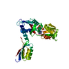
Mass: 96.063 Da / Num. of mol.: 3 / Source method: obtained synthetically / Formula: SO4
#3: Chemical ChemComp-MG / MAGNESIUM ION


Mass: 24.305 Da / Num. of mol.: 2 / Source method: obtained synthetically / Formula: Mg
#4: Chemical ChemComp-ATP / ADENOSINE-5'-TRIPHOSPHATE


Mass: 507.181 Da / Num. of mol.: 1 / Source method: obtained synthetically / Formula: C10H16N5O13P3 / Comment: ATP, energy-carrying molecule*YM
#5: Water ChemComp-HOH / water


Mass: 18.015 Da / Num. of mol.: 28 / Source method: isolated from a natural source / Formula: H2O

-
Experimental details

-
Experiment

ExperimentMethod: X-RAY DIFFRACTION / Number of used crystals: 1

-
Sample preparation

CrystalDensity Matthews: 3.16 Å3/Da / Density % sol: 61.09 % / Description: Crystals grew as medium sizes prisms overnight
Crystal growTemperature: 293.15 K / Method: vapor diffusion, hanging drop / pH: 6.5
Details: 1.6 M magnesium sulphate and 0.1 M, 2-(N-morpholino)ethanesulfonic acid

-
Data collection

DiffractionMean temperature: 100 K
Diffraction sourceSource: SYNCHROTRON / Site: Australian Synchrotron / Beamline: MX1 / Wavelength: 0.9537 Å
DetectorType: ADSC QUANTUM 210r / Detector: CCD / Date: Mar 25, 2015
RadiationProtocol: SINGLE WAVELENGTH / Monochromatic (M) / Laue (L): M / Scattering type: x-ray
Radiation wavelengthWavelength: 0.9537 Å / Relative weight: 1
ReflectionResolution: 2.4→80.43 Å / Num. obs: 15289 / % possible obs: 100 % / Redundancy: 20 % / CC1/2: 0.773 / Rmerge(I) obs: 0.143 / Rpim(I) all: 0.492 / Net I/σ(I): 20

-
Processing

Software
NameVersionClassification
REFMAC5.8.0158refinement
XDSdata reduction
Aimlessdata scaling
PHASERphasing
RefinementMethod to determine structure: MOLECULAR REPLACEMENT
Starting model: PDB:1NH7
Resolution: 2.4→80.43 Å / Cor.coef. Fo:Fc: 0.944 / Cor.coef. Fo:Fc free: 0.934 / SU B: 16.133 / SU ML: 0.198 / Cross valid method: THROUGHOUT / ESU R: 0.308 / ESU R Free: 0.232 / Stereochemistry target values: MAXIMUM LIKELIHOOD / Details: HYDROGENS HAVE BEEN ADDED IN THE RIDING POSITIONS
RfactorNum. reflection% reflectionSelection details
Rfree0.25051 747 4.9 %RANDOM
Rwork0.21571 ---
obs0.21749 14540 99.96 %-
Solvent computationIon probe radii: 0.8 Å / Shrinkage radii: 0.8 Å / VDW probe radii: 1.2 Å / Solvent model: MASK
Displacement parametersBiso mean: 55.545 Å2
Baniso -1Baniso -2Baniso -3
1-1.64 Å20.82 Å2-0 Å2
2--1.64 Å2-0 Å2
3----5.33 Å2
Refinement stepCycle: 1 / Resolution: 2.4→80.43 Å
ProteinNucleic acidLigandSolventTotal
Num. atoms1981 0 48 28 2057
Refine LS restraints
Refine-IDTypeDev idealDev ideal targetNumber
X-RAY DIFFRACTIONr_bond_refined_d0.010.0192096
X-RAY DIFFRACTIONr_bond_other_d0.0020.021924
X-RAY DIFFRACTIONr_angle_refined_deg1.5192.0032873
X-RAY DIFFRACTIONr_angle_other_deg0.95834400
X-RAY DIFFRACTIONr_dihedral_angle_1_deg6.9295279
X-RAY DIFFRACTIONr_dihedral_angle_2_deg40.63723.37874
X-RAY DIFFRACTIONr_dihedral_angle_3_deg14.88115288
X-RAY DIFFRACTIONr_dihedral_angle_4_deg21.0341515
X-RAY DIFFRACTIONr_chiral_restr0.080.2347
X-RAY DIFFRACTIONr_gen_planes_refined0.0050.0212358
X-RAY DIFFRACTIONr_gen_planes_other0.0010.02420
X-RAY DIFFRACTIONr_nbd_refined
X-RAY DIFFRACTIONr_nbd_other
X-RAY DIFFRACTIONr_nbtor_refined
X-RAY DIFFRACTIONr_nbtor_other
X-RAY DIFFRACTIONr_xyhbond_nbd_refined
X-RAY DIFFRACTIONr_xyhbond_nbd_other
X-RAY DIFFRACTIONr_metal_ion_refined
X-RAY DIFFRACTIONr_metal_ion_other
X-RAY DIFFRACTIONr_symmetry_vdw_refined
X-RAY DIFFRACTIONr_symmetry_vdw_other
X-RAY DIFFRACTIONr_symmetry_hbond_refined
X-RAY DIFFRACTIONr_symmetry_hbond_other
X-RAY DIFFRACTIONr_symmetry_metal_ion_refined
X-RAY DIFFRACTIONr_symmetry_metal_ion_other
X-RAY DIFFRACTIONr_mcbond_it1.1413.5861122
X-RAY DIFFRACTIONr_mcbond_other1.1413.5851121
X-RAY DIFFRACTIONr_mcangle_it1.985.3731399
X-RAY DIFFRACTIONr_mcangle_other1.9795.3751400
X-RAY DIFFRACTIONr_scbond_it1.3033.761973
X-RAY DIFFRACTIONr_scbond_other1.3023.761974
X-RAY DIFFRACTIONr_scangle_it
X-RAY DIFFRACTIONr_scangle_other2.0635.6151475
X-RAY DIFFRACTIONr_long_range_B_refined4.40142.8112153
X-RAY DIFFRACTIONr_long_range_B_other4.38942.7852151
X-RAY DIFFRACTIONr_rigid_bond_restr
X-RAY DIFFRACTIONr_sphericity_free
X-RAY DIFFRACTIONr_sphericity_bonded
LS refinement shellResolution: 2.4→2.463 Å / Total num. of bins used: 20
RfactorNum. reflection% reflection
Rfree0.357 61 -
Rwork0.317 1069 -
obs--100 %
Refinement TLS params.Method: refined / Origin x: -21.69 Å / Origin y: 4.5235 Å / Origin z: -36.1812 Å
111213212223313233
T0.0876 Å20.0031 Å20.0901 Å2-0.1263 Å2-0.0047 Å2--0.1406 Å2
L1.4725 °20.0761 °20.7157 °2-1.1684 °20.1385 °2--1.6532 °2
S0.1187 Å °-0.2718 Å °-0.0119 Å °0.1218 Å °0.007 Å °0.1194 Å °-0.1513 Å °-0.2673 Å °-0.1257 Å °

+
About Yorodumi

-
News

-
Feb 9, 2022. New format data for meta-information of EMDB entries

New format data for meta-information of EMDB entries

  • Version 3 of the EMDB header file is now the official format.
  • The previous official version 1.9 will be removed from the archive.

Related info.:EMDB header

External links:wwPDB to switch to version 3 of the EMDB data model

-
Aug 12, 2020. Covid-19 info

Covid-19 info

URL: https://pdbjlvh1.pdbj.org/emnavi/covid19.php

New page: Covid-19 featured information page in EM Navigator.

Related info.:Covid-19 info / Mar 5, 2020. Novel coronavirus structure data

+
Mar 5, 2020. Novel coronavirus structure data

Novel coronavirus structure data

Related info.:Yorodumi Speices / Aug 12, 2020. Covid-19 info

External links:COVID-19 featured content - PDBj / Molecule of the Month (242):Coronavirus Proteases

+
Jan 31, 2019. EMDB accession codes are about to change! (news from PDBe EMDB page)

EMDB accession codes are about to change! (news from PDBe EMDB page)

  • The allocation of 4 digits for EMDB accession codes will soon come to an end. Whilst these codes will remain in use, new EMDB accession codes will include an additional digit and will expand incrementally as the available range of codes is exhausted. The current 4-digit format prefixed with “EMD-” (i.e. EMD-XXXX) will advance to a 5-digit format (i.e. EMD-XXXXX), and so on. It is currently estimated that the 4-digit codes will be depleted around Spring 2019, at which point the 5-digit format will come into force.
  • The EM Navigator/Yorodumi systems omit the EMD- prefix.

Related info.:Q: What is EMD? / ID/Accession-code notation in Yorodumi/EM Navigator

External links:EMDB Accession Codes are Changing Soon! / Contact to PDBj

+
Jul 12, 2017. Major update of PDB

Major update of PDB

  • wwPDB released updated PDB data conforming to the new PDBx/mmCIF dictionary.
  • This is a major update changing the version number from 4 to 5, and with Remediation, in which all the entries are updated.
  • In this update, many items about electron microscopy experimental information are reorganized (e.g. em_software).
  • Now, EM Navigator and Yorodumi are based on the updated data.

External links:wwPDB Remediation / Enriched Model Files Conforming to OneDep Data Standards Now Available in the PDB FTP Archive

-
Yorodumi

Thousand views of thousand structures

  • Yorodumi is a browser for structure data from EMDB, PDB, SASBDB, etc.
  • This page is also the successor to EM Navigator detail page, and also detail information page/front-end page for Omokage search.
  • The word "yorodu" (or yorozu) is an old Japanese word meaning "ten thousand". "mi" (miru) is to see.

Related info.:EMDB / PDB / SASBDB / Comparison of 3 databanks / Yorodumi Search / Aug 31, 2016. New EM Navigator & Yorodumi / Yorodumi Papers / Jmol/JSmol / Function and homology information / Changes in new EM Navigator and Yorodumi

Read more