 Movie
Movie Controller

[English] 日本語
 Yorodumi
- PDB-2f3m: Structure of human GLUTATHIONE S-TRANSFERASE M1A-1A complexed wit... -
+ Open data

- Basic information
| Entry | Database: PDB / ID: 2f3m | ||||||
|---|---|---|---|---|---|---|---|
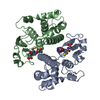
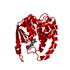
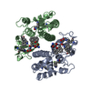
| Title | Structure of human GLUTATHIONE S-TRANSFERASE M1A-1A complexed with 1-(S-(GLUTATHIONYL)-2,4,6-TRINITROCYCLOHEXADIENATE ANION | ||||||
 Components Components | Glutathione S-transferase Mu 1 | ||||||
 Keywords Keywords | TRANSFERASE / GLUTATHIONE / CONJUGATION / DETOXIFICATION / CYTOSOLIC / DIMER / ACTIVE SITE / TRANSITION STATE ANALOGUE | ||||||
| Function / homology |  Function and homology information Function and homology informationnitrobenzene metabolic process / cellular detoxification of nitrogen compound / hepoxilin biosynthetic process / glutathione derivative biosynthetic process / glutathione binding / Glutathione conjugation / Paracetamol ADME / Azathioprine ADME / prostaglandin metabolic process / glutathione transferase ...nitrobenzene metabolic process / cellular detoxification of nitrogen compound / hepoxilin biosynthetic process / glutathione derivative biosynthetic process / glutathione binding / Glutathione conjugation / Paracetamol ADME / Azathioprine ADME / prostaglandin metabolic process / glutathione transferase / glutathione transferase activity / xenobiotic catabolic process / glutathione metabolic process / enzyme binding / protein homodimerization activity / cytosol / cytoplasm Similarity search - Function | ||||||
| Biological species |  Homo sapiens (human) Homo sapiens (human) | ||||||
| Method |  X-RAY DIFFRACTION / MOLECULAR REPLACEMENT / Resolution: 2.7 Å X-RAY DIFFRACTION / MOLECULAR REPLACEMENT / Resolution: 2.7 Å | ||||||
 Authors Authors | Patskovsky, Y. / Patskovska, L. / Almo, S.C. / Listowsky, I. | ||||||
 Citation Citation | Journal: Biochemistry / Year: 2006 Title: Transition state model and mechanism of nucleophilic aromatic substitution reactions catalyzed by human glutathione S-transferase M1a-1a. Authors: Patskovsky, Y. / Patskovska, L. / Almo, S.C. / Listowsky, I. #1: Journal: Biochemistry / Year: 1999Title: Function of His107 in the Catalytic Mechanism of Human Glutathione S-Transferase Hgstm1A-1A Authors: Patskovsky, Y.V. / Patskovska, L.N. / Listowsky, I. | ||||||
| History |
|
- Structure visualization
| Structure viewer | Molecule: MolmilJmol/JSmol |
|---|
- Downloads & links
-Download
| PDBx/mmCIF format | 2f3m.cif.gz | 275.1 KB | Display | PDBx/mmCIF format |
|---|---|---|---|---|
| PDB format | pdb2f3m.ent.gz | 224.5 KB | Display | PDB format |
| PDBx/mmJSON format | 2f3m.json.gz | Tree view | PDBx/mmJSON format | |
| Others |  Other downloads Other downloads |
-Validation report
| Arichive directory | https://data.pdbj.org/pub/pdb/validation_reports/f3/2f3mftp://data.pdbj.org/pub/pdb/validation_reports/f3/2f3m | HTTPS FTP |
|---|
-Related structure data
| Related structure data | 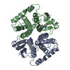 1xw6C  1xwkC  1gtuS S: Starting model for refinement C: citing same article ( |
|---|---|
| Similar structure data |


-Links
 PDBj
PDBj

- Assembly
| Deposited unit | 
| ||||||||||||||||||||||||||||||||||||||||||
|---|---|---|---|---|---|---|---|---|---|---|---|---|---|---|---|---|---|---|---|---|---|---|---|---|---|---|---|---|---|---|---|---|---|---|---|---|---|---|---|---|---|---|---|
| 1 | 
| ||||||||||||||||||||||||||||||||||||||||||
| 2 | 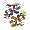
| ||||||||||||||||||||||||||||||||||||||||||
| 3 | 
| ||||||||||||||||||||||||||||||||||||||||||
| Unit cell |
| ||||||||||||||||||||||||||||||||||||||||||
| Noncrystallographic symmetry (NCS) | NCS domain:
NCS domain segments: Component-ID: 1 / Ens-ID: 1 / Beg auth comp-ID: PRO / Beg label comp-ID: PRO / End auth comp-ID: LYS / End label comp-ID: LYS / Refine code: 1 / Auth seq-ID: 1 - 217 / Label seq-ID: 2 - 218
| ||||||||||||||||||||||||||||||||||||||||||
| Details | The biological assembly is a homodimer composed of two identical monomers. The unit cell/asymmetric unit contains 3 full homodimers |
-Components
| #1: Protein | Mass: 25746.842 Da / Num. of mol.: 6 Source method: isolated from a genetically manipulated source Source: (gene. exp.) Homo sapiens (human) / Gene: GSTM1, GST1 / Plasmid: pET3a / Species (production host): Escherichia coli / Production host:  #2: Chemical | ChemComp-GTD /   Mass: 520.428 Da / Num. of mol.: 6 / Source method: obtained synthetically / Formula: C16H20N6O12S Mass: 520.428 Da / Num. of mol.: 6 / Source method: obtained synthetically / Formula: C16H20N6O12S#3: Water | ChemComp-HOH / |  Mass: 18.015 Da / Num. of mol.: 53 / Source method: isolated from a natural source / Formula: H2O Mass: 18.015 Da / Num. of mol.: 53 / Source method: isolated from a natural source / Formula: H2OHas protein modification | Y | |
|---|
-Experimental details
-Experiment
| Experiment | Method: X-RAY DIFFRACTION / Number of used crystals: 1 |
|---|
- Sample preparation
| Crystal | Density Matthews: 2.35 Å3/Da / Density % sol: 47.15 % |
|---|---|
| Crystal grow | Temperature: 280 K / pH: 6 Details: 20% PEG 4000, pH 6.00, VAPOR DIFFUSION, temperature 280K |
-Data collection
| Diffraction | Mean temperature: 100 K |
|---|---|
| Diffraction source | Source: ROTATING ANODE / Type: RIGAKU RU200 / Wavelength: 1.5418 |
| Detector | Type: RIGAKU RAXIS IV / Detector: IMAGE PLATE / Date: Nov 11, 2005 / Details: MIRRORS |
| Radiation | Monochromator: MIRRORS / Protocol: SINGLE WAVELENGTH / Monochromatic (M) / Laue (L): M / Scattering type: x-ray |
| Radiation wavelength | Wavelength: 1.5418 Å / Relative weight: 1 |
| Reflection | Resolution: 2.667→100 Å / Num. obs: 38744 / % possible obs: 97.1 % / Observed criterion σ(I): 0 / Redundancy: 1.9 % / Rmerge(I) obs: 0.068 / Rsym value: 0.042 / Net I/σ(I): 7.8 |
| Reflection shell | Resolution: 2.67→2.8 Å / Redundancy: 1.8 % / Rmerge(I) obs: 0.38 / Mean I/σ(I) obs: 1.7 / Rsym value: 0.32 / % possible all: 95.1 |
- Processing
| Software |
| ||||||||||||||||||||||||||||||||||||||||||||||||||||||||||||||||||||||||||||||||||||||||||||||||||||||||||||||||||||||||||||||||||||||||||||||||||||||||||||||||||||||||||
|---|---|---|---|---|---|---|---|---|---|---|---|---|---|---|---|---|---|---|---|---|---|---|---|---|---|---|---|---|---|---|---|---|---|---|---|---|---|---|---|---|---|---|---|---|---|---|---|---|---|---|---|---|---|---|---|---|---|---|---|---|---|---|---|---|---|---|---|---|---|---|---|---|---|---|---|---|---|---|---|---|---|---|---|---|---|---|---|---|---|---|---|---|---|---|---|---|---|---|---|---|---|---|---|---|---|---|---|---|---|---|---|---|---|---|---|---|---|---|---|---|---|---|---|---|---|---|---|---|---|---|---|---|---|---|---|---|---|---|---|---|---|---|---|---|---|---|---|---|---|---|---|---|---|---|---|---|---|---|---|---|---|---|---|---|---|---|---|---|---|---|---|
| Refinement | Method to determine structure: MOLECULAR REPLACEMENT Starting model: 1GTU Resolution: 2.7→20 Å / SU B: 20.024 / SU ML: 0.381 / Cross valid method: THROUGHOUT / σ(F): 0 / ESU R Free: 0.402 / Stereochemistry target values: MAXIMUM LIKELIHOOD / Details: HYDROGENS HAVE BEEN ADDED IN THE RIDING POSITIONS
| ||||||||||||||||||||||||||||||||||||||||||||||||||||||||||||||||||||||||||||||||||||||||||||||||||||||||||||||||||||||||||||||||||||||||||||||||||||||||||||||||||||||||||
| Solvent computation | Ion probe radii: 0.8 Å / Shrinkage radii: 0.8 Å / VDW probe radii: 1.2 Å / Solvent model: MASK | ||||||||||||||||||||||||||||||||||||||||||||||||||||||||||||||||||||||||||||||||||||||||||||||||||||||||||||||||||||||||||||||||||||||||||||||||||||||||||||||||||||||||||
| Displacement parameters | Biso mean: 54.38 Å2
| ||||||||||||||||||||||||||||||||||||||||||||||||||||||||||||||||||||||||||||||||||||||||||||||||||||||||||||||||||||||||||||||||||||||||||||||||||||||||||||||||||||||||||
| Refine analyze | Luzzati coordinate error obs: 0.44 Å | ||||||||||||||||||||||||||||||||||||||||||||||||||||||||||||||||||||||||||||||||||||||||||||||||||||||||||||||||||||||||||||||||||||||||||||||||||||||||||||||||||||||||||
| Refinement step | Cycle: LAST / Resolution: 2.7→20 Å
| ||||||||||||||||||||||||||||||||||||||||||||||||||||||||||||||||||||||||||||||||||||||||||||||||||||||||||||||||||||||||||||||||||||||||||||||||||||||||||||||||||||||||||
| Refine LS restraints |
| ||||||||||||||||||||||||||||||||||||||||||||||||||||||||||||||||||||||||||||||||||||||||||||||||||||||||||||||||||||||||||||||||||||||||||||||||||||||||||||||||||||||||||
| Refine LS restraints NCS | Ens-ID: 1 / Number: 1789 / Refine-ID: X-RAY DIFFRACTION
| ||||||||||||||||||||||||||||||||||||||||||||||||||||||||||||||||||||||||||||||||||||||||||||||||||||||||||||||||||||||||||||||||||||||||||||||||||||||||||||||||||||||||||
| LS refinement shell | Resolution: 2.7→2.77 Å / Total num. of bins used: 20
|
