 Movie
Movie Controller


+ Open data

- Basic information
| Entry | Database: PDB / ID: 1gtu | ||||||
|---|---|---|---|---|---|---|---|
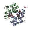
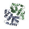
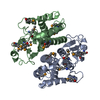
| Title | LIGAND-FREE HUMAN GLUTATHIONE S-TRANSFERASE M1A-1A | ||||||
 Components Components | GLUTATHIONE S-TRANSFERASE | ||||||
 Keywords Keywords | TRANSFERASE / GLUTATHIONE / CONJUGATION / DETOXIFICATION / CYTOSOLIC / DIMER | ||||||
| Function / homology |  Function and homology information Function and homology informationnitrobenzene metabolic process / cellular detoxification of nitrogen compound / hepoxilin biosynthetic process / glutathione derivative biosynthetic process / glutathione binding / Glutathione conjugation / Paracetamol ADME / Azathioprine ADME / prostaglandin metabolic process / glutathione transferase ...nitrobenzene metabolic process / cellular detoxification of nitrogen compound / hepoxilin biosynthetic process / glutathione derivative biosynthetic process / glutathione binding / Glutathione conjugation / Paracetamol ADME / Azathioprine ADME / prostaglandin metabolic process / glutathione transferase / glutathione transferase activity / xenobiotic catabolic process / glutathione metabolic process / enzyme binding / protein homodimerization activity / cytoplasm / cytosol Similarity search - Function | ||||||
| Biological species |  Homo sapiens (human) Homo sapiens (human) | ||||||
| Method |  X-RAY DIFFRACTION / MOLECULAR REPLACEMENT / Resolution: 2.68 Å X-RAY DIFFRACTION / MOLECULAR REPLACEMENT / Resolution: 2.68 Å | ||||||
 Authors Authors | Patskovsky, Y.V. / Patskovska, L.N. / Listowsky, I. | ||||||
 Citation Citation | Journal: Biochemistry / Year: 1999 Title: Functions of His107 in the catalytic mechanism of human glutathione S-transferase hGSTM1a-1a. Authors: Patskovsky, Y.V. / Patskovska, L.N. / Listowsky, I. #1: Journal: Biochem.J. / Year: 1993Title: Deduced Amino Acid Sequence, Gene Structure and Chromosomal Location of a Novel Human Class Mu Glutathione S-Transferase, Gstm4 Authors: Zhong, S. / Spurr, N.K. / Hayes, J.D. / Wolf, C.R. #2: Journal: Proc.Natl.Acad.Sci.USA / Year: 1988Title: Hereditary Differences in the Expression of the Human Glutathione Transferase Active on Trans-Stilbene Oxide are due to a Gene Deletion Authors: Seidegard, J. / Vorachek, W.R. / Pero, R.W. / Pearson, W.R. #3: Journal: Nucleic Acids Res. / Year: 1988Title: The Human Liver Glutathione S-Transferase Gene Superfamily: Expression and Chromosome Mapping of an Hb Subunit Cdna Authors: Dejong, J.L. / Chang, C.M. / Whang-Peng, J. / Knutsen, T. / TU, C.P. | ||||||
| History |
|
- Structure visualization
| Structure viewer | Molecule: MolmilJmol/JSmol |
|---|
- Downloads & links
-Download
| PDBx/mmCIF format | 1gtu.cif.gz | 182.6 KB | Display | PDBx/mmCIF format |
|---|---|---|---|---|
| PDB format | pdb1gtu.ent.gz | 149.3 KB | Display | PDB format |
| PDBx/mmJSON format | 1gtu.json.gz | Tree view | PDBx/mmJSON format | |
| Others |  Other downloads Other downloads |
-Validation report
| Arichive directory | https://data.pdbj.org/pub/pdb/validation_reports/gt/1gtuftp://data.pdbj.org/pub/pdb/validation_reports/gt/1gtu | HTTPS FTP |
|---|
-Related structure data
| Related structure data | 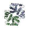 2gtuS S: Starting model for refinement |
|---|---|
| Similar structure data |


-Links
 PDBj
PDBj

- Assembly
| Deposited unit | 
| ||||||||||||||||
|---|---|---|---|---|---|---|---|---|---|---|---|---|---|---|---|---|---|
| 1 | 
| ||||||||||||||||
| 2 | 
| ||||||||||||||||
| Unit cell |
| ||||||||||||||||
| Noncrystallographic symmetry (NCS) | NCS oper:
|
-Components
| #1: Protein | Mass: 25615.646 Da / Num. of mol.: 4 Source method: isolated from a genetically manipulated source Details: LIGAND-FREE / Source: (gene. exp.) Homo sapiens (human)Description: THE GSTM1A CDNA WAS AMPLIFIED USING RT-PCR AND SUBCLONED INTO A PET3A EXPRESSION VECTOR Cell line: HELA / Cellular location: CYTOPLASM / Gene: GSTM1A / Organ: LIVER / Plasmid: PET3A-GSTM1A / Species (production host): Escherichia coli / Gene (production host): GSTM1A / Production host:  #2: Water | ChemComp-HOH / |  Mass: 18.015 Da / Num. of mol.: 63 / Source method: isolated from a natural source / Formula: H2O Mass: 18.015 Da / Num. of mol.: 63 / Source method: isolated from a natural source / Formula: H2O |
|---|
-Experimental details
-Experiment
| Experiment | Method: X-RAY DIFFRACTION / Number of used crystals: 2 |
|---|
- Sample preparation
| Crystal | Density Matthews: 2.87 Å3/Da / Density % sol: 58 % | ||||||||||||||||||||||||||||||||||||||||||
|---|---|---|---|---|---|---|---|---|---|---|---|---|---|---|---|---|---|---|---|---|---|---|---|---|---|---|---|---|---|---|---|---|---|---|---|---|---|---|---|---|---|---|---|
| Crystal grow | pH: 6 / Details: pH 6.0 | ||||||||||||||||||||||||||||||||||||||||||
| Crystal grow | *PLUS Temperature: 289 K / pH: 7.5 / Method: vapor diffusion, hanging drop | ||||||||||||||||||||||||||||||||||||||||||
| Components of the solutions | *PLUS
|
-Data collection
| Diffraction | Mean temperature: 290 K |
|---|---|
| Diffraction source | Source: ROTATING ANODE / Type: RIGAKU RUH2R / Wavelength: 1.5418 |
| Detector | Type: SIEMENS-NICOLET X100 / Detector: AREA DETECTOR / Date: Apr 1, 1997 |
| Radiation | Monochromator: GRAPHITE(002) / Monochromatic (M) / Laue (L): M / Scattering type: x-ray |
| Radiation wavelength | Wavelength: 1.5418 Å / Relative weight: 1 |
| Reflection | Resolution: 2.68→12.35 Å / Num. obs: 26217 / % possible obs: 98 % / Observed criterion σ(I): 2 / Redundancy: 2 % / Rmerge(I) obs: 0.058 |
| Reflection shell | Resolution: 2.68→2.9 Å / Redundancy: 1.75 % / Rmerge(I) obs: 0.155 / % possible all: 87.1 |
| Reflection shell | *PLUS % possible obs: 87.1 % |
- Processing
| Software |
| ||||||||||||||||||||||||||||||||||||||||||||||||||||||||||||||||||||||||||||||||
|---|---|---|---|---|---|---|---|---|---|---|---|---|---|---|---|---|---|---|---|---|---|---|---|---|---|---|---|---|---|---|---|---|---|---|---|---|---|---|---|---|---|---|---|---|---|---|---|---|---|---|---|---|---|---|---|---|---|---|---|---|---|---|---|---|---|---|---|---|---|---|---|---|---|---|---|---|---|---|---|---|---|
| Refinement | Method to determine structure: MOLECULAR REPLACEMENT Starting model: PDB ENTRY 2GTU Resolution: 2.68→10 Å / Rfactor Rfree error: 0.008 / Data cutoff high absF: 1000000 / Data cutoff low absF: 0.001 / Isotropic thermal model: RESTRAINED / Cross valid method: THROUGHOUT / σ(F): 2
| ||||||||||||||||||||||||||||||||||||||||||||||||||||||||||||||||||||||||||||||||
| Refine analyze |
| ||||||||||||||||||||||||||||||||||||||||||||||||||||||||||||||||||||||||||||||||
| Refinement step | Cycle: LAST / Resolution: 2.68→10 Å
| ||||||||||||||||||||||||||||||||||||||||||||||||||||||||||||||||||||||||||||||||
| Refine LS restraints |
| ||||||||||||||||||||||||||||||||||||||||||||||||||||||||||||||||||||||||||||||||
| LS refinement shell | Resolution: 2.68→2.8 Å / Rfactor Rfree error: 0.01 / Total num. of bins used: 8
| ||||||||||||||||||||||||||||||||||||||||||||||||||||||||||||||||||||||||||||||||
| Xplor file |
| ||||||||||||||||||||||||||||||||||||||||||||||||||||||||||||||||||||||||||||||||
| Software | *PLUS Name: X-PLOR / Version: 3.851 / Classification: refinement | ||||||||||||||||||||||||||||||||||||||||||||||||||||||||||||||||||||||||||||||||
| Refine LS restraints | *PLUS
|
