 Movie
Movie Controller


+ Open data

- Basic information
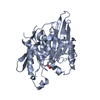
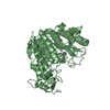
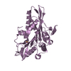
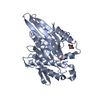
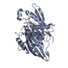
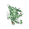
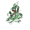
| Entry | Database: PDB / ID: 2f2e | ||||||
|---|---|---|---|---|---|---|---|
| Title | Crystal Structure of PA1607, a Putative Transcription Factor | ||||||
 Components Components | PA1607 | ||||||
 Keywords Keywords | DNA BINDING PROTEIN/STRUCTURAL GENOMICS / Transcription factor / helix-trun-helix / APC5613 / structural genomics / PSI / Protein Structure Initiative / Midwest Center for Structural Genomics / MCSG / DNA BINDING PROTEIN-STRUCTURAL GENOMICS COMPLEX | ||||||
| Function / homology |  Function and homology information Function and homology informationprotein-DNA complex / DNA-binding transcription factor activity / regulation of DNA-templated transcription / DNA-templated transcription / DNA binding Similarity search - Function | ||||||
| Biological species |   Pseudomonas aeruginosa (bacteria) Pseudomonas aeruginosa (bacteria) | ||||||
| Method |  X-RAY DIFFRACTION / SYNCHROTRON / MAD / Resolution: 1.85 Å X-RAY DIFFRACTION / SYNCHROTRON / MAD / Resolution: 1.85 Å | ||||||
 Authors Authors | Sieminska, E.A. / Xu, X. / Zheng, H. / Lunin, V. / Cuff, M. / Joachimiak, A. / Edwards, A. / Savchenko, A. / Sanders, D.A. / Midwest Center for Structural Genomics (MCSG) | ||||||
 Citation Citation | Journal: Protein Sci. / Year: 2007 Title: The X-ray crystal structure of PA1607 from Pseudomonas aureginosa at 1.9 A resolution--a putative transcription factor. Authors: Sieminska, E.A. / Xu, X. / Savchenko, A. / Sanders, D.A. | ||||||
| History |
|
- Structure visualization
| Structure viewer | Molecule: MolmilJmol/JSmol |
|---|
- Downloads & links
-Download
| PDBx/mmCIF format | 2f2e.cif.gz | 77.8 KB | Display | PDBx/mmCIF format |
|---|---|---|---|---|
| PDB format | pdb2f2e.ent.gz | 58.3 KB | Display | PDB format |
| PDBx/mmJSON format | 2f2e.json.gz | Tree view | PDBx/mmJSON format | |
| Others |  Other downloads Other downloads |
-Validation report
| Arichive directory | https://data.pdbj.org/pub/pdb/validation_reports/f2/2f2eftp://data.pdbj.org/pub/pdb/validation_reports/f2/2f2e | HTTPS FTP |
|---|
-Related structure data
| Similar structure data | |
|---|---|
| Other databases |


-Links
 PDBj
PDBj

- Assembly
| Deposited unit | 
| |||||||||
|---|---|---|---|---|---|---|---|---|---|---|
| 1 |
| |||||||||
| 2 | 
| |||||||||
| 3 | 
| |||||||||
| Unit cell |
| |||||||||
| Components on special symmetry positions |
| |||||||||
| Details | This protein is a dimer in solution |
-Components
| #1: Protein | Mass: 16373.173 Da / Num. of mol.: 2 Source method: isolated from a genetically manipulated source Source: (gene. exp.) Pseudomonas aeruginosa (bacteria) / Gene: PA1607 / Plasmid: pET15-modified / Production host: #2: Chemical | ChemComp-SO4 /   Mass: 96.063 Da / Num. of mol.: 4 / Source method: obtained synthetically / Formula: SO4 Mass: 96.063 Da / Num. of mol.: 4 / Source method: obtained synthetically / Formula: SO4#3: Sugar | ChemComp-GLC / |   Type: D-saccharide, alpha linking / Mass: 180.156 Da / Num. of mol.: 1 Type: D-saccharide, alpha linking / Mass: 180.156 Da / Num. of mol.: 1Source method: isolated from a genetically manipulated source Formula: C6H12O6 #4: Water | ChemComp-HOH / |  Mass: 18.015 Da / Num. of mol.: 329 / Source method: isolated from a natural source / Formula: H2O Mass: 18.015 Da / Num. of mol.: 329 / Source method: isolated from a natural source / Formula: H2OHas protein modification | Y | |
|---|
-Experimental details
-Experiment
| Experiment | Method: X-RAY DIFFRACTION / Number of used crystals: 1 |
|---|
- Sample preparation
| Crystal | Density Matthews: 2.23 Å3/Da / Density % sol: 44.75 % |
|---|---|
| Crystal grow | Temperature: 298 K / Method: vapor diffusion, hanging drop / pH: 8.6 Details: 10mg/ml protein, 0.1 M Tris, 0.2 M LiSO4, 28 % PEG 3350, pH 8.6, VAPOR DIFFUSION, HANGING DROP, temperature 298K |
-Data collection
| Diffraction source | Source: SYNCHROTRON / Site: APS  / Beamline: 19-ID / Wavelength: 0.97932, 0.97948 / Beamline: 19-ID / Wavelength: 0.97932, 0.97948 | |||||||||
|---|---|---|---|---|---|---|---|---|---|---|
| Detector | Date: Apr 1, 2005 | |||||||||
| Radiation | Protocol: MAD / Monochromatic (M) / Laue (L): M / Scattering type: x-ray | |||||||||
| Radiation wavelength |
| |||||||||
| Reflection | Resolution: 1.85→78 Å / Num. obs: 24599 |
- Processing
| Software |
| ||||||||||||||||||||||||||||||||||||||||||||||||||||||||||||||||||||||||||||||||||||||||||||||||||||||||||||||||||||||||||||||||||||||||||||||||||||||||||||||||||||||||||
|---|---|---|---|---|---|---|---|---|---|---|---|---|---|---|---|---|---|---|---|---|---|---|---|---|---|---|---|---|---|---|---|---|---|---|---|---|---|---|---|---|---|---|---|---|---|---|---|---|---|---|---|---|---|---|---|---|---|---|---|---|---|---|---|---|---|---|---|---|---|---|---|---|---|---|---|---|---|---|---|---|---|---|---|---|---|---|---|---|---|---|---|---|---|---|---|---|---|---|---|---|---|---|---|---|---|---|---|---|---|---|---|---|---|---|---|---|---|---|---|---|---|---|---|---|---|---|---|---|---|---|---|---|---|---|---|---|---|---|---|---|---|---|---|---|---|---|---|---|---|---|---|---|---|---|---|---|---|---|---|---|---|---|---|---|---|---|---|---|---|---|---|
| Refinement | Method to determine structure: MAD / Resolution: 1.85→35.27 Å / Cor.coef. Fo:Fc: 0.96 / Cor.coef. Fo:Fc free: 0.919 / SU B: 6.596 / SU ML: 0.109 / Cross valid method: THROUGHOUT / ESU R: 0.167 / ESU R Free: 0.17 / Stereochemistry target values: MAXIMUM LIKELIHOOD
| ||||||||||||||||||||||||||||||||||||||||||||||||||||||||||||||||||||||||||||||||||||||||||||||||||||||||||||||||||||||||||||||||||||||||||||||||||||||||||||||||||||||||||
| Solvent computation | Ion probe radii: 0.8 Å / Shrinkage radii: 0.8 Å / VDW probe radii: 1.2 Å / Solvent model: MASK | ||||||||||||||||||||||||||||||||||||||||||||||||||||||||||||||||||||||||||||||||||||||||||||||||||||||||||||||||||||||||||||||||||||||||||||||||||||||||||||||||||||||||||
| Displacement parameters | Biso mean: 36.558 Å2
| ||||||||||||||||||||||||||||||||||||||||||||||||||||||||||||||||||||||||||||||||||||||||||||||||||||||||||||||||||||||||||||||||||||||||||||||||||||||||||||||||||||||||||
| Refinement step | Cycle: LAST / Resolution: 1.85→35.27 Å
| ||||||||||||||||||||||||||||||||||||||||||||||||||||||||||||||||||||||||||||||||||||||||||||||||||||||||||||||||||||||||||||||||||||||||||||||||||||||||||||||||||||||||||
| Refine LS restraints |
| ||||||||||||||||||||||||||||||||||||||||||||||||||||||||||||||||||||||||||||||||||||||||||||||||||||||||||||||||||||||||||||||||||||||||||||||||||||||||||||||||||||||||||
| LS refinement shell | Resolution: 1.85→1.897 Å / Total num. of bins used: 20
|
