 Movie
Movie Controller

[English] 日本語
 Yorodumi
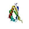
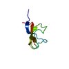
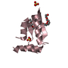
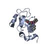
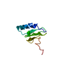
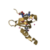
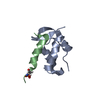
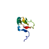
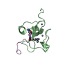
- PDB-2evb: Structure of Biotin Carboxyl Carrier Protein (74Val start) from P... -
+ Open data

- Basic information
| Entry | Database: PDB / ID: 2evb | ||||||
|---|---|---|---|---|---|---|---|
| Title | Structure of Biotin Carboxyl Carrier Protein (74Val start) from Pyrococcus horikoshi OT3 Ligand Free Form I | ||||||
 Components Components | methylmalonyl-CoA decarboxylase gamma chain | ||||||
 Keywords Keywords | LIPID BINDING PROTEIN / TRANSFERASE / Biotin / BCCP / Structural Genomics / NPPSFA / National Project on Protein Structural and Functional Analyses / RIKEN Structural Genomics/Proteomics Initiative / RSGI | ||||||
| Function / homology |  Function and homology information Function and homology information: / Biotin-binding site / Biotin-requiring enzymes attachment site. / RNA polymerase II/Efflux pump adaptor protein, barrel-sandwich hybrid domain / Biotin-requiring enzyme / Biotinyl/lipoyl domain profile. / Biotin/lipoyl attachment / Single hybrid motif / OB fold (Dihydrolipoamide Acetyltransferase, E2P) / Beta Barrel / Mainly Beta Similarity search - Domain/homology | ||||||
| Biological species |   Pyrococcus horikoshii (archaea) Pyrococcus horikoshii (archaea) | ||||||
| Method |  X-RAY DIFFRACTION / SYNCHROTRON / MOLECULAR REPLACEMENT / Resolution: 1.55 Å X-RAY DIFFRACTION / SYNCHROTRON / MOLECULAR REPLACEMENT / Resolution: 1.55 Å | ||||||
 Authors Authors | Bagautdinov, B. / Kunishima, N. / RIKEN Structural Genomics/Proteomics Initiative (RSGI) | ||||||
 Citation Citation | Journal: J.Biol.Chem. / Year: 2008 Title: Protein biotinylation visualized by a complex structure of biotin protein ligase with a substrate Authors: Bagautdinov, B. / Matsuura, Y. / Bagautdinova, S. / Kunishima, N. | ||||||
| History |
|
- Structure visualization
| Structure viewer | Molecule: MolmilJmol/JSmol |
|---|
- Downloads & links
-Download
| PDBx/mmCIF format | 2evb.cif.gz | 27.6 KB | Display | PDBx/mmCIF format |
|---|---|---|---|---|
| PDB format | pdb2evb.ent.gz | 17.6 KB | Display | PDB format |
| PDBx/mmJSON format | 2evb.json.gz | Tree view | PDBx/mmJSON format | |
| Others |  Other downloads Other downloads |
-Validation report
| Arichive directory | https://data.pdbj.org/pub/pdb/validation_reports/ev/2evbftp://data.pdbj.org/pub/pdb/validation_reports/ev/2evb | HTTPS FTP |
|---|
-Related structure data
| Related structure data |  1x01C  2d5dC 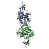 2dxuC  2dzcC  2e41C  2e64C  2ejfC  2ejgC  2zgwC 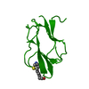 1bdoS C: citing same article ( S: Starting model for refinement |
|---|---|
| Similar structure data | |
| Other databases |

-Links
 PDBj
PDBj

- Assembly
| Deposited unit | 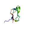
| ||||||||
|---|---|---|---|---|---|---|---|---|---|
| 1 |
| ||||||||
| Unit cell |
|
-Components
| #1: Protein | Mass: 7985.457 Da / Num. of mol.: 1 / Fragment: residues 73-146 Source method: isolated from a genetically manipulated source Source: (gene. exp.) Pyrococcus horikoshii (archaea) / Strain: OT3 / Gene: bccp / Plasmid: PET 11A / Species (production host): Escherichia coli / Production host:  References: UniProt: O59021, methylmalonyl-CoA carboxytransferase |
|---|---|
| #2: Water | ChemComp-HOH /  Mass: 18.015 Da / Num. of mol.: 111 / Source method: isolated from a natural source / Formula: H2O Mass: 18.015 Da / Num. of mol.: 111 / Source method: isolated from a natural source / Formula: H2O |
-Experimental details
-Experiment
| Experiment | Method: X-RAY DIFFRACTION / Number of used crystals: 1 |
|---|
- Sample preparation
| Crystal | Density Matthews: 1.931508 Å3/Da / Density % sol: 36.319191 % |
|---|---|
| Crystal grow | Temperature: 295 K / Method: microbatch / pH: 4.62 Details: PEG 550 MME 55w/v(%), Acetate, NaOH , pH 4.62, microbatch, temperature 295K |
-Data collection
| Diffraction | Mean temperature: 100 K |
|---|---|
| Diffraction source | Source: SYNCHROTRON / Site: SPring-8  / Beamline: BL26B1 / Wavelength: 1 Å / Beamline: BL26B1 / Wavelength: 1 Å |
| Detector | Type: MARRESEARCH / Detector: CCD / Date: Oct 2, 2005 / Details: mirrors |
| Radiation | Monochromator: GRAPHITE / Protocol: SINGLE WAVELENGTH / Monochromatic (M) / Laue (L): M / Scattering type: x-ray |
| Radiation wavelength | Wavelength: 1 Å / Relative weight: 1 |
| Reflection | Resolution: 1.55→24.5 Å / Num. all: 8858 / Num. obs: 8821 / % possible obs: 93.3 % / Observed criterion σ(F): 0 / Observed criterion σ(I): 0 / Redundancy: 5.7 % / Biso Wilson estimate: 23.82 Å2 / Rmerge(I) obs: 0.051 / Rsym value: 0.05 / Net I/σ(I): 22.3 |
| Reflection shell | Resolution: 1.55→1.61 Å / Redundancy: 2.9 % / Rmerge(I) obs: 0.195 / Mean I/σ(I) obs: 0.165 / Num. unique all: 560 / % possible all: 62.2 |
- Processing
| Software |
| |||||||||||||||||||||||||
|---|---|---|---|---|---|---|---|---|---|---|---|---|---|---|---|---|---|---|---|---|---|---|---|---|---|---|
| Refinement | Method to determine structure: MOLECULAR REPLACEMENT Starting model: PDB ENTRY 1BDO Resolution: 1.55→24.5 Å / Isotropic thermal model: OVERALL / Cross valid method: THROUGHOUT / σ(F): 0 / σ(I): 0 / Stereochemistry target values: Engh & Huber
| |||||||||||||||||||||||||
| Displacement parameters | Biso mean: 30.2 Å2
| |||||||||||||||||||||||||
| Refine analyze |
| |||||||||||||||||||||||||
| Refinement step | Cycle: LAST / Resolution: 1.55→24.5 Å
| |||||||||||||||||||||||||
| Refine LS restraints |
| |||||||||||||||||||||||||
| LS refinement shell | Refine-ID: X-RAY DIFFRACTION / Total num. of bins used: 2
|
