 Movie
Movie Controller


+ Open data

- Basic information
| Entry | Database: PDB / ID: 2ebo | ||||||
|---|---|---|---|---|---|---|---|
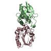
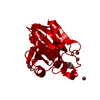
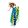
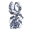
| Title | CORE STRUCTURE OF GP2 FROM EBOLA VIRUS | ||||||
 Components Components | EBOLA VIRUS ENVELOPE GLYCOPROTEIN | ||||||
 Keywords Keywords | ENVELOPE GLYCOPROTEIN / FILOVIRUS / EBOLA VIRUS / GP2 / COAT PROTEIN | ||||||
| Function / homology |  Function and homology information Function and homology informationsymbiont-mediated killing of host cell / host cell endoplasmic reticulum / viral budding from plasma membrane / symbiont-mediated-mediated suppression of host tetherin activity / clathrin-dependent endocytosis of virus by host cell / host cell cytoplasm / entry receptor-mediated virion attachment to host cell / symbiont-mediated suppression of host innate immune response / membrane raft / fusion of virus membrane with host endosome membrane ...symbiont-mediated killing of host cell / host cell endoplasmic reticulum / viral budding from plasma membrane / symbiont-mediated-mediated suppression of host tetherin activity / clathrin-dependent endocytosis of virus by host cell / host cell cytoplasm / entry receptor-mediated virion attachment to host cell / symbiont-mediated suppression of host innate immune response / membrane raft / fusion of virus membrane with host endosome membrane / viral envelope / symbiont entry into host cell / lipid binding / host cell plasma membrane / virion membrane / extracellular region / identical protein binding Similarity search - Function | ||||||
| Biological species | Zaire ebolavirus - Mayinga (Zaire 1976) | ||||||
| Method |  X-RAY DIFFRACTION / SYNCHROTRON / MOLECULAR REPLACEMENT / Resolution: 1.9 Å X-RAY DIFFRACTION / SYNCHROTRON / MOLECULAR REPLACEMENT / Resolution: 1.9 Å | ||||||
 Authors Authors | Malashkevich, V.N. / Schneider, B.J. / Mcnally, M.L. / Milhollen, M.A. / Pang, J.X. / Kim, P.S. | ||||||
 Citation Citation | Journal: Proc.Natl.Acad.Sci.USA / Year: 1999 Title: Core structure of the envelope glycoprotein GP2 from Ebola virus at 1.9-A resolution. Authors: Malashkevich, V.N. / Schneider, B.J. / McNally, M.L. / Milhollen, M.A. / Pang, J.X. / Kim, P.S. #1: Journal: Proc.Natl.Acad.Sci.USA / Year: 1998Title: Crystal Structure of the Simian Immunodeficiency Virus (Siv) Gp41 Core: Conserved Helical Interactions Underlie the Broad Inhibitory Activity of Gp41 Peptides Authors: Malashkevich, V.N. / Chan, D.C. / Chutkowski, C.T. / Kim, P.S. #2: Journal: Cell(Cambridge,Mass.) / Year: 1997Title: Core Structure of Gp41 from the HIV Envelope Glycoprotein Authors: Chan, D.C. / Fass, D. / Berger, J.M. / Kim, P.S. | ||||||
| History |
|
- Structure visualization
| Structure viewer | Molecule: MolmilJmol/JSmol |
|---|
- Downloads & links
-Download
| PDBx/mmCIF format | 2ebo.cif.gz | 60.9 KB | Display | PDBx/mmCIF format |
|---|---|---|---|---|
| PDB format | pdb2ebo.ent.gz | 45.6 KB | Display | PDB format |
| PDBx/mmJSON format | 2ebo.json.gz | Tree view | PDBx/mmJSON format | |
| Others |  Other downloads Other downloads |
-Validation report
| Arichive directory | https://data.pdbj.org/pub/pdb/validation_reports/eb/2eboftp://data.pdbj.org/pub/pdb/validation_reports/eb/2ebo | HTTPS FTP |
|---|
-Related structure data
| Related structure data |  1mofS S: Starting model for refinement |
|---|---|
| Similar structure data |


-Links
 PDBj
PDBj

- Assembly
| Deposited unit | 
| ||||||||||||
|---|---|---|---|---|---|---|---|---|---|---|---|---|---|
| 1 |
| ||||||||||||
| Unit cell |
| ||||||||||||
| Noncrystallographic symmetry (NCS) | NCS oper:
|
-Components
| #1: Protein | Mass: 8544.757 Da / Num. of mol.: 3 / Fragment: PROTEASE-RESISTANT FRAGMENT / Mutation: C609A Source method: isolated from a genetically manipulated source Source: (gene. exp.)   Zaire ebolavirus - Mayinga (Zaire, 1976) Zaire ebolavirus - Mayinga (Zaire, 1976)Genus: Ebola-like viruses / Species: Zaire ebolavirus / Strain: Zaire Mayinga / Cellular location: VIRAL MEMBRANE / Gene: GP41 / Variant: ZAIRE ISOLATE / Plasmid: PMMHB / Species (production host): Escherichia coli / Gene (production host): GP41 / Production host:  #2: Chemical | ChemComp-CL / |   Mass: 35.453 Da / Num. of mol.: 1 / Source method: obtained synthetically / Formula: Cl Mass: 35.453 Da / Num. of mol.: 1 / Source method: obtained synthetically / Formula: Cl#3: Water | ChemComp-HOH / |  Mass: 18.015 Da / Num. of mol.: 214 / Source method: isolated from a natural source / Formula: H2O Mass: 18.015 Da / Num. of mol.: 214 / Source method: isolated from a natural source / Formula: H2OHas protein modification | Y | |
|---|
-Experimental details
-Experiment
| Experiment | Method: X-RAY DIFFRACTION / Number of used crystals: 1 |
|---|
- Sample preparation
| Crystal | Density Matthews: 2.2 Å3/Da / Density % sol: 44 % Description: TRIAL MODEL CONTAINED ONLY 36% OF THE ATOMS OF THE TARGET STRUCTURE. | |||||||||||||||||||||||||||||||||||
|---|---|---|---|---|---|---|---|---|---|---|---|---|---|---|---|---|---|---|---|---|---|---|---|---|---|---|---|---|---|---|---|---|---|---|---|---|
| Crystal grow | pH: 4.6 / Details: pH 4.6 | |||||||||||||||||||||||||||||||||||
| Crystal grow | *PLUS Method: vapor diffusion, hanging drop | |||||||||||||||||||||||||||||||||||
| Components of the solutions | *PLUS
|
-Data collection
| Diffraction | Mean temperature: 100 K |
|---|---|
| Diffraction source | Source: SYNCHROTRON / Site: ALS  / Beamline: 5.0.2 / Wavelength: 1.01 / Beamline: 5.0.2 / Wavelength: 1.01 |
| Detector | Type: ADSC / Detector: CCD / Date: Dec 1, 1998 |
| Radiation | Monochromatic (M) / Laue (L): M / Scattering type: x-ray |
| Radiation wavelength | Wavelength: 1.01 Å / Relative weight: 1 |
| Reflection | Resolution: 1.9→25 Å / Num. obs: 17326 / % possible obs: 98.9 % / Observed criterion σ(I): 0 / Redundancy: 4.9 % / Biso Wilson estimate: 27.2 Å2 / Rmerge(I) obs: 0.037 / Rsym value: 0.037 / Net I/σ(I): 18.8 |
| Reflection shell | Resolution: 1.9→1.92 Å / Redundancy: 4 % / Rmerge(I) obs: 0.309 / Mean I/σ(I) obs: 3.5 / Rsym value: 0.309 / % possible all: 99.1 |
| Reflection | *PLUS Highest resolution: 1.9 Å / Lowest resolution: 20 Å / Num. measured all: 86630 |
| Reflection shell | *PLUS Highest resolution: 1.9 Å / Lowest resolution: 1.93 Å / % possible obs: 99.1 % |
- Processing
| Software |
| ||||||||||||||||||||||||||||||||||||||||||||||||||||||||||||||||||||||||||||||||
|---|---|---|---|---|---|---|---|---|---|---|---|---|---|---|---|---|---|---|---|---|---|---|---|---|---|---|---|---|---|---|---|---|---|---|---|---|---|---|---|---|---|---|---|---|---|---|---|---|---|---|---|---|---|---|---|---|---|---|---|---|---|---|---|---|---|---|---|---|---|---|---|---|---|---|---|---|---|---|---|---|---|
| Refinement | Method to determine structure: MOLECULAR REPLACEMENT Starting model: PDB ENTRY 1MOF Resolution: 1.9→20 Å / Rfactor Rfree error: 0.006 / Data cutoff high absF: 1363158.21 / Data cutoff low absF: 0 / Isotropic thermal model: RESTRAINED / Cross valid method: THROUGHOUT / σ(F): 0
| ||||||||||||||||||||||||||||||||||||||||||||||||||||||||||||||||||||||||||||||||
| Solvent computation | Solvent model: FLAT MODEL / Bsol: 96.68 Å2 / ksol: 0.34 e/Å3 | ||||||||||||||||||||||||||||||||||||||||||||||||||||||||||||||||||||||||||||||||
| Displacement parameters | Biso mean: 40.9 Å2
| ||||||||||||||||||||||||||||||||||||||||||||||||||||||||||||||||||||||||||||||||
| Refine analyze |
| ||||||||||||||||||||||||||||||||||||||||||||||||||||||||||||||||||||||||||||||||
| Refinement step | Cycle: LAST / Resolution: 1.9→20 Å
| ||||||||||||||||||||||||||||||||||||||||||||||||||||||||||||||||||||||||||||||||
| Refine LS restraints |
| ||||||||||||||||||||||||||||||||||||||||||||||||||||||||||||||||||||||||||||||||
| LS refinement shell | Resolution: 1.9→2.02 Å / Rfactor Rfree error: 0.019 / Total num. of bins used: 6
| ||||||||||||||||||||||||||||||||||||||||||||||||||||||||||||||||||||||||||||||||
| Xplor file |
| ||||||||||||||||||||||||||||||||||||||||||||||||||||||||||||||||||||||||||||||||
| Refinement | *PLUS % reflection Rfree: 10 % / Rfactor Rfree: 0.269 | ||||||||||||||||||||||||||||||||||||||||||||||||||||||||||||||||||||||||||||||||
| Solvent computation | *PLUS | ||||||||||||||||||||||||||||||||||||||||||||||||||||||||||||||||||||||||||||||||
| Displacement parameters | *PLUS | ||||||||||||||||||||||||||||||||||||||||||||||||||||||||||||||||||||||||||||||||
| Refine LS restraints | *PLUS
| ||||||||||||||||||||||||||||||||||||||||||||||||||||||||||||||||||||||||||||||||
| LS refinement shell | *PLUS Lowest resolution: 1.93 Å / Rfactor obs: 0.239 |
