 Movie
Movie Controller

[English] 日本語
 Yorodumi
- PDB-1msb: STRUCTURE OF THE CALCIUM-DEPENDENT LECTIN DOMAIN FROM A RAT MANNO... -
+ Open data

- Basic information
| Entry | Database: PDB / ID: 1msb | ||||||
|---|---|---|---|---|---|---|---|
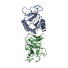
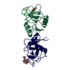
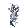
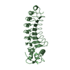
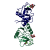
| Title | STRUCTURE OF THE CALCIUM-DEPENDENT LECTIN DOMAIN FROM A RAT MANNOSE-BINDING PROTEIN DETERMINED BY MAD PHASING | ||||||
 Components Components | MANNOSE-BINDING PROTEIN-A | ||||||
 Keywords Keywords | HEPATIC LECTIN | ||||||
| Function / homology |  Function and homology information Function and homology informationcalcium-dependent carbohydrate binding / complement activation, lectin pathway / oligosaccharide binding / : / surfactant homeostasis / phosphatidylinositol-4-phosphate binding / protein homotrimerization / D-mannose binding / polysaccharide binding / complement activation, classical pathway ...calcium-dependent carbohydrate binding / complement activation, lectin pathway / oligosaccharide binding / : / surfactant homeostasis / phosphatidylinositol-4-phosphate binding / protein homotrimerization / D-mannose binding / polysaccharide binding / complement activation, classical pathway / multivesicular body / positive regulation of phagocytosis / calcium-dependent protein binding / protease binding / defense response to Gram-positive bacterium / calcium ion binding / protein homodimerization activity / : / identical protein binding Similarity search - Function | ||||||
| Biological species |  | ||||||
| Method |  X-RAY DIFFRACTION / Resolution: 2.3 Å X-RAY DIFFRACTION / Resolution: 2.3 Å | ||||||
 Authors Authors | Weis, W.I. / Drickamer, K. / Hendrickson, W.A. | ||||||
 Citation Citation | Journal: Science / Year: 1991 Title: Structure of the calcium-dependent lectin domain from a rat mannose-binding protein determined by MAD phasing. Authors: Weis, W.I. / Kahn, R. / Fourme, R. / Drickamer, K. / Hendrickson, W.A. #1: Journal: J.Biol.Chem. / Year: 1991Title: Physical Characterization and Crystallization of the Carbohydrate-Recognition Domain of a Mannose-Binding Protein from Rat Authors: Weis, W.I. / Crichlow, G.V. / Murthy, H.M.K. / Hendrickson, W.A. / Drickamer, K. | ||||||
| History |
|
- Structure visualization
| Structure viewer | Molecule: MolmilJmol/JSmol |
|---|
- Downloads & links
-Download
| PDBx/mmCIF format | 1msb.cif.gz | 57.3 KB | Display | PDBx/mmCIF format |
|---|---|---|---|---|
| PDB format | pdb1msb.ent.gz | 41.8 KB | Display | PDB format |
| PDBx/mmJSON format | 1msb.json.gz | Tree view | PDBx/mmJSON format | |
| Others |  Other downloads Other downloads |
-Validation report
| Arichive directory | https://data.pdbj.org/pub/pdb/validation_reports/ms/1msbftp://data.pdbj.org/pub/pdb/validation_reports/ms/1msb | HTTPS FTP |
|---|
-Related structure data
| Similar structure data |
|---|


-Links
 PDBj
PDBj


- Assembly
| Deposited unit | 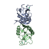
| ||||||||
|---|---|---|---|---|---|---|---|---|---|
| 1 |
| ||||||||
| Unit cell |
| ||||||||
| Atom site foot note | 1: RESIDUES PRO A 186 AND PRO B 186 ARE CIS PROLINES. 2: THE FIRST TWO RESIDUES OF CHAIN A, THE FIRST THREE RESIDUES OF CHAIN B, AND THE LAST 2 RESIDUES OF CHAIN B ARE NOT WELL DEFINED. | ||||||||
| Noncrystallographic symmetry (NCS) | NCS oper: (Code: given Matrix: (0.219746, -0.318844, 0.921982), Vector: |
-Components
| #1: Protein | Mass: 12688.150 Da / Num. of mol.: 2 Source method: isolated from a genetically manipulated source Source: (gene. exp.) #2: Chemical | ChemComp-HO /   Mass: 164.930 Da / Num. of mol.: 4 / Source method: obtained synthetically / Formula: Ho Mass: 164.930 Da / Num. of mol.: 4 / Source method: obtained synthetically / Formula: Ho#3: Water | ChemComp-HOH / |  Mass: 18.015 Da / Num. of mol.: 57 / Source method: isolated from a natural source / Formula: H2O Mass: 18.015 Da / Num. of mol.: 57 / Source method: isolated from a natural source / Formula: H2OHas protein modification | Y | |
|---|
-Experimental details
-Experiment
| Experiment | Method: X-RAY DIFFRACTION |
|---|
- Sample preparation
| Crystal | Density Matthews: 2.17 Å3/Da / Density % sol: 43.2 % | ||||||||||||||||||||||||||||||||||||||||||||||||||||||||||||||||||||||
|---|---|---|---|---|---|---|---|---|---|---|---|---|---|---|---|---|---|---|---|---|---|---|---|---|---|---|---|---|---|---|---|---|---|---|---|---|---|---|---|---|---|---|---|---|---|---|---|---|---|---|---|---|---|---|---|---|---|---|---|---|---|---|---|---|---|---|---|---|---|---|---|
| Crystal grow | *PLUS pH: 8 / Method: unknown | ||||||||||||||||||||||||||||||||||||||||||||||||||||||||||||||||||||||
| Components of the solutions | *PLUS
|
-Data collection
| Radiation | Scattering type: x-ray |
|---|---|
| Radiation wavelength | Relative weight: 1 |
| Reflection | *PLUS Num. obs: 7195 / % possible obs: 89.3 % / Rmerge(I) obs: 0.031 |
- Processing
| Software | Name: X-PLOR / Classification: refinement | ||||||||||||||||||||||||||||||||||||||||||||||||||||||||||||
|---|---|---|---|---|---|---|---|---|---|---|---|---|---|---|---|---|---|---|---|---|---|---|---|---|---|---|---|---|---|---|---|---|---|---|---|---|---|---|---|---|---|---|---|---|---|---|---|---|---|---|---|---|---|---|---|---|---|---|---|---|---|
| Refinement | Rfactor Rwork: 0.176 / Highest resolution: 2.3 Å Details: THE FIRST TWO RESIDUES OF CHAIN A, THE FIRST THREE RESIDUES OF CHAIN B, AND THE LAST 2 RESIDUES OF CHAIN B ARE NOT WELL DEFINED. | ||||||||||||||||||||||||||||||||||||||||||||||||||||||||||||
| Refinement step | Cycle: LAST / Highest resolution: 2.3 Å
| ||||||||||||||||||||||||||||||||||||||||||||||||||||||||||||
| Refine LS restraints |
| ||||||||||||||||||||||||||||||||||||||||||||||||||||||||||||
| Refinement | *PLUS Highest resolution: 2.3 Å / Num. reflection all: 1839 | ||||||||||||||||||||||||||||||||||||||||||||||||||||||||||||
| Solvent computation | *PLUS | ||||||||||||||||||||||||||||||||||||||||||||||||||||||||||||
| Displacement parameters | *PLUS | ||||||||||||||||||||||||||||||||||||||||||||||||||||||||||||
| Refine LS restraints | *PLUS Type: x_angle_d |
