 Movie
Movie Controller

[English] 日本語
 Yorodumi
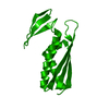
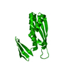
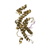
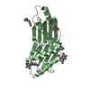
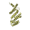
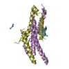
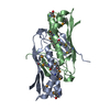
- PDB-2e8e: Crystal Structure of a hypothetical protein (Aq_1549) from Aquife... -
+ Open data

- Basic information
| Entry | Database: PDB / ID: 2e8e | ||||||
|---|---|---|---|---|---|---|---|
| Title | Crystal Structure of a hypothetical protein (Aq_1549) from Aquifex aeolicus VF5 (Reduced form) | ||||||
 Components Components | Hypothetical protein Aq_1549 | ||||||
 Keywords Keywords | STRUCTURAL GENOMICS / UNKNOWN FUNCTION / hypothetical protein / Aq_1549 / Osmatically induced protein / NPPSFA / National Project on Protein Structural and Functional Analyses / RIKEN Structural Genomics/Proteomics Initiative / RSGI | ||||||
| Function / homology |  Function and homology information Function and homology informationOsmC/Ohr family / OsmC/Ohr superfamily / OsmC-like protein / K homology (KH) domain / GMP Synthetase; Chain A, domain 3 / K homology domain-like, alpha/beta / 2-Layer Sandwich / Alpha Beta Similarity search - Domain/homology | ||||||
| Biological species |   Aquifex aeolicus (bacteria) Aquifex aeolicus (bacteria) | ||||||
| Method |  X-RAY DIFFRACTION / SYNCHROTRON / MOLECULAR REPLACEMENT / Resolution: 1.7 Å X-RAY DIFFRACTION / SYNCHROTRON / MOLECULAR REPLACEMENT / Resolution: 1.7 Å | ||||||
 Authors Authors | Kumarevel, T.S. / Kuramitsu, S. / Yokoyama, S. / RIKEN Structural Genomics/Proteomics Initiative (RSGI) | ||||||
 Citation Citation | Journal: To be Published Title: Crystal Structure of a hypothetical protein (Aq_1549) from Aquifex aeolicus VF5 Authors: Kumarevel, T.S. / Kuramitsu, S. / Yokoyama, S. | ||||||
| History |
|
- Structure visualization
| Structure viewer | Molecule: MolmilJmol/JSmol |
|---|
- Downloads & links
-Download
| PDBx/mmCIF format | 2e8e.cif.gz | 41.7 KB | Display | PDBx/mmCIF format |
|---|---|---|---|---|
| PDB format | pdb2e8e.ent.gz | 28 KB | Display | PDB format |
| PDBx/mmJSON format | 2e8e.json.gz | Tree view | PDBx/mmJSON format | |
| Others |  Other downloads Other downloads |
-Validation report
| Arichive directory | https://data.pdbj.org/pub/pdb/validation_reports/e8/2e8eftp://data.pdbj.org/pub/pdb/validation_reports/e8/2e8e | HTTPS FTP |
|---|
-Related structure data
| Related structure data |  2e8fC  2e8cS S: Starting model for refinement C: citing same article ( |
|---|---|
| Similar structure data | |
| Other databases |


-Links
 PDBj
PDBj


- Assembly
| Deposited unit | 
| ||||||||
|---|---|---|---|---|---|---|---|---|---|
| 1 | 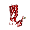
| ||||||||
| Unit cell |
|
-Components
| #1: Protein | Mass: 15037.618 Da / Num. of mol.: 1 Source method: isolated from a genetically manipulated source Source: (gene. exp.) Aquifex aeolicus (bacteria) / Strain: VF5 / Plasmid: pET21a / Production host: |
|---|---|
| #2: Chemical | ChemComp-DTT /   Mass: 154.251 Da / Num. of mol.: 1 / Source method: obtained synthetically / Formula: C4H10O2S2 Mass: 154.251 Da / Num. of mol.: 1 / Source method: obtained synthetically / Formula: C4H10O2S2 |
| #3: Water | ChemComp-HOH /  Mass: 18.015 Da / Num. of mol.: 91 / Source method: isolated from a natural source / Formula: H2O Mass: 18.015 Da / Num. of mol.: 91 / Source method: isolated from a natural source / Formula: H2O |
-Experimental details
-Experiment
| Experiment | Method: X-RAY DIFFRACTION / Number of used crystals: 1 |
|---|
- Sample preparation
| Crystal | Density Matthews: 2.14 Å3/Da / Density % sol: 42.51 % |
|---|---|
| Crystal grow | Temperature: 293 K / Method: vapor diffusion, sitting drop / pH: 5.5 Details: 50% PEG 200, 0.1M Citrate, pH 5.5, VAPOR DIFFUSION, SITTING DROP, temperature 293K |
-Data collection
| Diffraction | Mean temperature: 100 K |
|---|---|
| Diffraction source | Source: SYNCHROTRON / Site: SPring-8  / Beamline: BL26B1 / Wavelength: 1 Å / Beamline: BL26B1 / Wavelength: 1 Å |
| Detector | Type: ADSC QUANTUM 210 / Detector: CCD / Date: Sep 26, 2006 / Details: Si |
| Radiation | Protocol: SINGLE WAVELENGTH / Monochromatic (M) / Laue (L): M / Scattering type: x-ray |
| Radiation wavelength | Wavelength: 1 Å / Relative weight: 1 |
| Reflection | Resolution: 1.7→40 Å / Num. all: 14883 / Num. obs: 14883 / % possible obs: 99.9 % / Observed criterion σ(F): 2 / Observed criterion σ(I): 2 / Redundancy: 10 % / Biso Wilson estimate: 23.4 Å2 / Rmerge(I) obs: 0.064 |
| Reflection shell | Resolution: 1.7→1.76 Å / Redundancy: 10.1 % / Rmerge(I) obs: 0.367 / Num. unique all: 1450 / % possible all: 100 |
- Processing
| Software |
| ||||||||||||||||||||||||||||||||||||
|---|---|---|---|---|---|---|---|---|---|---|---|---|---|---|---|---|---|---|---|---|---|---|---|---|---|---|---|---|---|---|---|---|---|---|---|---|---|
| Refinement | Method to determine structure: MOLECULAR REPLACEMENT Starting model: PDB ENTRY 2E8C Resolution: 1.7→19.58 Å / Rfactor Rfree error: 0.009 / Data cutoff high absF: 1124848.21 / Data cutoff low absF: 0 / Isotropic thermal model: RESTRAINED / Cross valid method: THROUGHOUT / σ(F): 0 / Stereochemistry target values: Engh & Huber
| ||||||||||||||||||||||||||||||||||||
| Solvent computation | Solvent model: FLAT MODEL / Bsol: 63.5113 Å2 / ksol: 0.397908 e/Å3 | ||||||||||||||||||||||||||||||||||||
| Displacement parameters | Biso mean: 26.5 Å2
| ||||||||||||||||||||||||||||||||||||
| Refine analyze |
| ||||||||||||||||||||||||||||||||||||
| Refinement step | Cycle: LAST / Resolution: 1.7→19.58 Å
| ||||||||||||||||||||||||||||||||||||
| Refine LS restraints |
| ||||||||||||||||||||||||||||||||||||
| LS refinement shell | Resolution: 1.7→1.81 Å / Rfactor Rfree error: 0.03 / Total num. of bins used: 6
| ||||||||||||||||||||||||||||||||||||
| Xplor file |
|
