 Movie
Movie Controller

[English] 日本語
 Yorodumi
- PDB-2e6c: Crystal structure of the stationary phase survival protein SurE f... -
+ Open data

- Basic information
| Entry | Database: PDB / ID: 2e6c | ||||||
|---|---|---|---|---|---|---|---|
| Title | Crystal structure of the stationary phase survival protein SurE from Thermus thermophilus HB8 cocrystallized with manganese and AMP | ||||||
 Components Components | 5'-nucleotidase surE | ||||||
 Keywords Keywords | HYDROLASE / SurE protein / cocrystal structure with manganese ion and AMP | ||||||
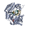
| Function / homology |  Function and homology information Function and homology information3'-nucleotidase activity / exopolyphosphatase activity / 5'-nucleotidase / 5'-nucleotidase activity / nucleotide binding / metal ion binding / cytoplasm Similarity search - Function | ||||||
| Biological species |   Thermus thermophilus (bacteria) Thermus thermophilus (bacteria) | ||||||
| Method |  X-RAY DIFFRACTION / SYNCHROTRON / MOLECULAR REPLACEMENT / Resolution: 2.05 Å X-RAY DIFFRACTION / SYNCHROTRON / MOLECULAR REPLACEMENT / Resolution: 2.05 Å | ||||||
 Authors Authors | Iwasaki, W. / Miki, K. | ||||||
 Citation Citation | Journal: J.Mol.Biol. / Year: 2007 Title: Crystal Structure of the Stationary Phase Survival Protein SurE with Metal Ion and AMP Authors: Iwasaki, W. / Miki, K. | ||||||
| History |
| ||||||
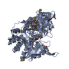
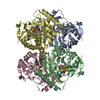
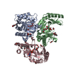
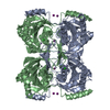
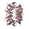
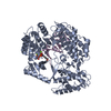
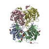
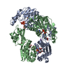
| Remark 300 | BIOMOLECULE 1 This entry contains the crystallographic asymmetric unit which consists of 4 chain(s) ...BIOMOLECULE 1 This entry contains the crystallographic asymmetric unit which consists of 4 chain(s) forming a tetramer. This protein is in a dimer-tetramer equilibrium in solution. It is unknown whether this protein is functional as a dimer or a tetramer. |
- Structure visualization
| Structure viewer | Molecule: MolmilJmol/JSmol |
|---|
- Downloads & links
-Download
| PDBx/mmCIF format | 2e6c.cif.gz | 200.1 KB | Display | PDBx/mmCIF format |
|---|---|---|---|---|
| PDB format | pdb2e6c.ent.gz | 159.7 KB | Display | PDB format |
| PDBx/mmJSON format | 2e6c.json.gz | Tree view | PDBx/mmJSON format | |
| Others |  Other downloads Other downloads |
-Validation report
| Arichive directory | https://data.pdbj.org/pub/pdb/validation_reports/e6/2e6cftp://data.pdbj.org/pub/pdb/validation_reports/e6/2e6c | HTTPS FTP |
|---|
-Related structure data
| Related structure data |  2e69SC  2e6bC  2e6eC  2e6gC  2e6hC S: Starting model for refinement C: citing same article ( |
|---|---|
| Similar structure data |


-Links
 PDBj
PDBj- Assembly
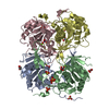
| Deposited unit | 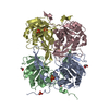
| ||||||||
|---|---|---|---|---|---|---|---|---|---|
| 1 |
| ||||||||
| Unit cell |
| ||||||||
| Details | probably tetramer |
-Components
| #1: Protein | Mass: 26625.287 Da / Num. of mol.: 4 Source method: isolated from a genetically manipulated source Source: (gene. exp.) Thermus thermophilus (bacteria) / Strain: HB8 / Plasmid: pET11a / Species (production host): Escherichia coli / Production host: #2: Chemical | ChemComp-MN /   Mass: 54.938 Da / Num. of mol.: 4 / Source method: obtained synthetically / Formula: Mn Mass: 54.938 Da / Num. of mol.: 4 / Source method: obtained synthetically / Formula: Mn#3: Chemical | ChemComp-SO4 /   Mass: 96.063 Da / Num. of mol.: 9 / Source method: obtained synthetically / Formula: SO4 Mass: 96.063 Da / Num. of mol.: 9 / Source method: obtained synthetically / Formula: SO4#4: Chemical |   Mass: 92.094 Da / Num. of mol.: 2 / Source method: obtained synthetically / Formula: C3H8O3 Mass: 92.094 Da / Num. of mol.: 2 / Source method: obtained synthetically / Formula: C3H8O3#5: Water | ChemComp-HOH / |  Mass: 18.015 Da / Num. of mol.: 304 / Source method: isolated from a natural source / Formula: H2O Mass: 18.015 Da / Num. of mol.: 304 / Source method: isolated from a natural source / Formula: H2O |
|---|
-Experimental details
-Experiment
| Experiment | Method: X-RAY DIFFRACTION / Number of used crystals: 1 |
|---|
- Sample preparation
| Crystal | Density Matthews: 3.11 Å3/Da / Density % sol: 60.5 % |
|---|---|
| Crystal grow | Temperature: 293 K / Method: vapor diffusion / pH: 8.5 Details: 0.1M Tris-HCl pH8.5, 0.75M ammonium sulfate, 10% glycerol, VAPOR DIFFUSION, temperature 293K |
-Data collection
| Diffraction | Mean temperature: 90 K |
|---|---|
| Diffraction source | Source: SYNCHROTRON / Site: SPring-8  / Beamline: BL44B2 / Wavelength: 1 Å / Beamline: BL44B2 / Wavelength: 1 Å |
| Detector | Type: ADSC QUANTUM 210 / Detector: CCD / Date: Jul 26, 2005 |
| Radiation | Protocol: SINGLE WAVELENGTH / Monochromatic (M) / Laue (L): M / Scattering type: x-ray |
| Radiation wavelength | Wavelength: 1 Å / Relative weight: 1 |
| Reflection | Resolution: 2.05→50 Å / Num. obs: 83555 / % possible obs: 100 % / Biso Wilson estimate: 18 Å2 / Rmerge(I) obs: 0.083 / Net I/σ(I): 20.3 |
| Reflection shell | Resolution: 2.05→2.12 Å / Rmerge(I) obs: 0.352 / Mean I/σ(I) obs: 7.5 / % possible all: 100 |
- Processing
| Software |
| ||||||||||||||||||||||||||||||||||||
|---|---|---|---|---|---|---|---|---|---|---|---|---|---|---|---|---|---|---|---|---|---|---|---|---|---|---|---|---|---|---|---|---|---|---|---|---|---|
| Refinement | Method to determine structure: MOLECULAR REPLACEMENT Starting model: PDB entry 2E69 Resolution: 2.05→46.56 Å / Rfactor Rfree error: 0.004 / Data cutoff high absF: 3382480.74 / Data cutoff low absF: 0 / Isotropic thermal model: RESTRAINED / Cross valid method: THROUGHOUT / σ(F): 0 / Stereochemistry target values: Engh & Huber
| ||||||||||||||||||||||||||||||||||||
| Solvent computation | Solvent model: FLAT MODEL / Bsol: 48.5791 Å2 / ksol: 0.374002 e/Å3 | ||||||||||||||||||||||||||||||||||||
| Displacement parameters | Biso mean: 32.3 Å2
| ||||||||||||||||||||||||||||||||||||
| Refine analyze |
| ||||||||||||||||||||||||||||||||||||
| Refinement step | Cycle: LAST / Resolution: 2.05→46.56 Å
| ||||||||||||||||||||||||||||||||||||
| Refine LS restraints |
| ||||||||||||||||||||||||||||||||||||
| LS refinement shell | Resolution: 2.05→2.18 Å / Rfactor Rfree error: 0.011 / Total num. of bins used: 6
| ||||||||||||||||||||||||||||||||||||
| Xplor file |
|
