| 登録情報 | データベース: PDB / ID: 2e4w
|
|---|
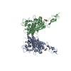
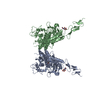
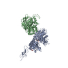
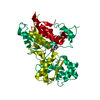
| タイトル | Crystal structure of the extracellular region of the group II metabotropic glutamate receptor complexed with 1S,3S-ACPD |
|---|
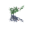
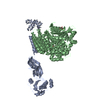
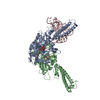
 要素 要素 | Metabotropic glutamate receptor 3 |
|---|
 キーワード キーワード | SIGNALING PROTEIN / G-PROTEIN-COUPLED RECEPTOR / NEURON / CENTRAL NERVE SYSTEM |
|---|
| 機能・相同性 |  機能・相同性情報 機能・相同性情報
Class C/3 (Metabotropic glutamate/pheromone receptors) / group II metabotropic glutamate receptor activity / G protein-coupled glutamate receptor signaling pathway / G alpha (i) signalling events / astrocyte projection / cellular response to stress / postsynaptic modulation of chemical synaptic transmission / regulation of synaptic transmission, glutamatergic / sensory perception of pain / calcium channel regulator activity ...Class C/3 (Metabotropic glutamate/pheromone receptors) / group II metabotropic glutamate receptor activity / G protein-coupled glutamate receptor signaling pathway / G alpha (i) signalling events / astrocyte projection / cellular response to stress / postsynaptic modulation of chemical synaptic transmission / regulation of synaptic transmission, glutamatergic / sensory perception of pain / calcium channel regulator activity / modulation of chemical synaptic transmission / adenylate cyclase-inhibiting G protein-coupled receptor signaling pathway / presynaptic membrane / scaffold protein binding / gene expression / dendritic spine / postsynaptic membrane / neuron projection / postsynaptic density / axon / glutamatergic synapse / plasma membrane類似検索 - 分子機能 GPCR, family 3, nine cysteines domain / GPCR, family 3, metabotropic glutamate receptor 3 / Tumor Necrosis Factor Receptor, subunit A; domain 2 / GPCR, family 3, metabotropic glutamate receptor / : / G-protein coupled receptors family 3 signature 1. / G-protein coupled receptors family 3 signature 3. / G-protein coupled receptors family 3 signature 2. / GPCR, family 3, nine cysteines domain / GPCR, family 3, nine cysteines domain superfamily ...GPCR, family 3, nine cysteines domain / GPCR, family 3, metabotropic glutamate receptor 3 / Tumor Necrosis Factor Receptor, subunit A; domain 2 / GPCR, family 3, metabotropic glutamate receptor / : / G-protein coupled receptors family 3 signature 1. / G-protein coupled receptors family 3 signature 3. / G-protein coupled receptors family 3 signature 2. / GPCR, family 3, nine cysteines domain / GPCR, family 3, nine cysteines domain superfamily / Nine Cysteines Domain of family 3 GPCR / GPCR, family 3, conserved site / GPCR, family 3 / G-protein coupled receptors family 3 profile. / GPCR family 3, C-terminal / 7 transmembrane sweet-taste receptor of 3 GCPR / Response regulator / Receptor, ligand binding region / Receptor family ligand binding region / Ribbon / Periplasmic binding protein-like I / Rossmann fold / 3-Layer(aba) Sandwich / Mainly Beta / Alpha Beta類似検索 - ドメイン・相同性 (1S,3S)-1-aminocyclopentane-1,3-dicarboxylic acid / Metabotropic glutamate receptor 3類似検索 - 構成要素 |
|---|
| 生物種 |   Rattus norvegicus (ドブネズミ) Rattus norvegicus (ドブネズミ) |
|---|
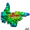
| 手法 |  X線回折 / シンクロトロン / 分子置換 / 解像度: 2.4 Å X線回折 / シンクロトロン / 分子置換 / 解像度: 2.4 Å |
|---|
 データ登録者 データ登録者 | Muto, T. / Tsuchiya, D. / Morikawa, K. / Jingami, H. |
|---|
 引用 引用 | ジャーナル: Proc.Natl.Acad.Sci.Usa / 年: 2007
タイトル: Structures of the extracellular regions of the group II/III metabotropic glutamate receptors
著者: Muto, T. / Tsuchiya, D. / Morikawa, K. / Jingami, H. |
|---|
| 履歴 | | 登録 | 2006年12月17日 | 登録サイト: PDBJ / 処理サイト: PDBJ |
|---|
| 改定 1.0 | 2007年2月27日 | Provider: repository / タイプ: Initial release |
|---|
| 改定 1.1 | 2008年4月30日 | Group: Version format compliance |
|---|
| 改定 1.2 | 2011年7月13日 | Group: Non-polymer description / Version format compliance |
|---|
| 改定 1.3 | 2020年7月29日 | Group: Data collection / Database references ...Data collection / Database references / Derived calculations / Structure summary
カテゴリ: chem_comp / entity ...chem_comp / entity / pdbx_chem_comp_identifier / pdbx_entity_nonpoly / struct_conn / struct_ref_seq_dif / struct_site / struct_site_gen
Item: _chem_comp.name / _chem_comp.type ..._chem_comp.name / _chem_comp.type / _entity.pdbx_description / _pdbx_entity_nonpoly.name / _struct_conn.pdbx_leaving_atom_flag / _struct_conn.pdbx_role / _struct_conn.ptnr1_auth_comp_id / _struct_conn.ptnr1_auth_seq_id / _struct_conn.ptnr1_label_asym_id / _struct_conn.ptnr1_label_atom_id / _struct_conn.ptnr1_label_comp_id / _struct_conn.ptnr1_label_seq_id / _struct_conn.ptnr2_auth_comp_id / _struct_conn.ptnr2_auth_seq_id / _struct_conn.ptnr2_label_asym_id / _struct_conn.ptnr2_label_atom_id / _struct_conn.ptnr2_label_comp_id / _struct_conn.ptnr2_label_seq_id / _struct_ref_seq_dif.details
解説: Carbohydrate remediation / Provider: repository / タイプ: Remediation |
|---|
| 改定 1.4 | 2021年11月10日 | Group: Database references / Structure summary / カテゴリ: chem_comp / database_2 / struct_ref_seq_dif
Item: _chem_comp.pdbx_synonyms / _database_2.pdbx_DOI ..._chem_comp.pdbx_synonyms / _database_2.pdbx_DOI / _database_2.pdbx_database_accession / _struct_ref_seq_dif.details |
|---|
| 改定 1.5 | 2023年10月25日 | Group: Data collection / Refinement description
カテゴリ: chem_comp_atom / chem_comp_bond / pdbx_initial_refinement_model |
|---|
| 改定 1.6 | 2024年10月23日 | Group: Structure summary
カテゴリ: pdbx_entry_details / pdbx_modification_feature |
|---|
|
|---|
 ムービー
ムービー コントローラー


 データを開く
 基本情報
基本情報 構造の表示
構造の表示 ダウンロードとリンク
 その他のダウンロード


 PDBj
PDBj


 集合体
集合体
 Trichoplusia ni (イラクサキンウワバ) / 参照: UniProt: P31422
 タイプ: D-saccharide, beta linking / 分子量: 221.208 Da / 分子数: 2 / 由来タイプ: 組換発現 / 式: C8H15NO6
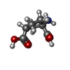
 分子量: 173.167 Da / 分子数: 2 / 由来タイプ: 合成 / 式: C7H11NO4
 分子量: 18.015 Da / 分子数: 295 / 由来タイプ: 天然 / 式: H2O
 試料調製
試料調製 / ビームライン: AR-NW12A / 波長: 1 Å
 解析
解析