 Movie
Movie Controller

[English] 日本語
 Yorodumi
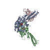
- PDB-2e4u: Crystal structure of the extracellular region of the group II met... -
+ Open data

- Basic information
| Entry | Database: PDB / ID: 2e4u | ||||||
|---|---|---|---|---|---|---|---|
| Title | Crystal structure of the extracellular region of the group II metabotropic glutamate receptor complexed with L-glutamate | ||||||
 Components Components | Metabotropic glutamate receptor 3 | ||||||
 Keywords Keywords | SIGNALING PROTEIN / G-protein-coupled receptor / neuron / central nerve system | ||||||
| Function / homology |  Function and homology information Function and homology informationClass C/3 (Metabotropic glutamate/pheromone receptors) / group II metabotropic glutamate receptor activity / G protein-coupled glutamate receptor signaling pathway / G alpha (i) signalling events / astrocyte projection / cellular response to stress / postsynaptic modulation of chemical synaptic transmission / regulation of synaptic transmission, glutamatergic / sensory perception of pain / calcium channel regulator activity ...Class C/3 (Metabotropic glutamate/pheromone receptors) / group II metabotropic glutamate receptor activity / G protein-coupled glutamate receptor signaling pathway / G alpha (i) signalling events / astrocyte projection / cellular response to stress / postsynaptic modulation of chemical synaptic transmission / regulation of synaptic transmission, glutamatergic / sensory perception of pain / calcium channel regulator activity / modulation of chemical synaptic transmission / adenylate cyclase-inhibiting G protein-coupled receptor signaling pathway / presynaptic membrane / scaffold protein binding / gene expression / dendritic spine / postsynaptic membrane / neuron projection / postsynaptic density / axon / glutamatergic synapse / plasma membrane Similarity search - Function | ||||||
| Biological species |  | ||||||
| Method |  X-RAY DIFFRACTION / SYNCHROTRON / MOLECULAR REPLACEMENT / Resolution: 2.35 Å X-RAY DIFFRACTION / SYNCHROTRON / MOLECULAR REPLACEMENT / Resolution: 2.35 Å | ||||||
 Authors Authors | Muto, T. / Tsuchiya, D. / Morikawa, K. / Jingami, H. | ||||||
 Citation Citation | Journal: Proc.Natl.Acad.Sci.Usa / Year: 2007 Title: Structures of the extracellular regions of the group II/III metabotropic glutamate receptors Authors: Muto, T. / Tsuchiya, D. / Morikawa, K. / Jingami, H. | ||||||
| History |
|
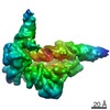
- Structure visualization
| Structure viewer | Molecule: MolmilJmol/JSmol |
|---|
- Downloads & links
-Download
| PDBx/mmCIF format | 2e4u.cif.gz | 220.1 KB | Display | PDBx/mmCIF format |
|---|---|---|---|---|
| PDB format | pdb2e4u.ent.gz | 174.1 KB | Display | PDB format |
| PDBx/mmJSON format | 2e4u.json.gz | Tree view | PDBx/mmJSON format | |
| Others |  Other downloads Other downloads |
-Validation report
| Arichive directory | https://data.pdbj.org/pub/pdb/validation_reports/e4/2e4uftp://data.pdbj.org/pub/pdb/validation_reports/e4/2e4u | HTTPS FTP |
|---|
-Related structure data
| Related structure data | 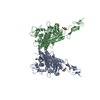 2e4vC 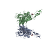 2e4wC 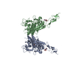 2e4xC 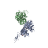 2e4yC 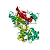 2e4zC  1ewkS C: citing same article ( S: Starting model for refinement |
|---|---|
| Similar structure data |


-Links
 PDBj
PDBj


- Assembly
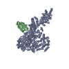
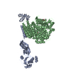
| Deposited unit | 
| ||||||||
|---|---|---|---|---|---|---|---|---|---|
| 1 |
| ||||||||
| Unit cell |
| ||||||||
| Details | The asymmetric unit contains one biological homodimer. |
-Components
| #1: Protein | Mass: 63117.297 Da / Num. of mol.: 2 / Fragment: Extracellular region / Mutation: N414Q, N439Q Source method: isolated from a genetically manipulated source Source: (gene. exp.)  Trichoplusia ni (cabbage looper) / References: UniProt: P31422 Trichoplusia ni (cabbage looper) / References: UniProt: P31422#2: Sugar |   Type: D-saccharide, beta linking / Mass: 221.208 Da / Num. of mol.: 2 Type: D-saccharide, beta linking / Mass: 221.208 Da / Num. of mol.: 2Source method: isolated from a genetically manipulated source Formula: C8H15NO6 #3: Chemical |   Type: L-peptide linking / Mass: 147.129 Da / Num. of mol.: 2 / Source method: obtained synthetically / Formula: C5H9NO4 Type: L-peptide linking / Mass: 147.129 Da / Num. of mol.: 2 / Source method: obtained synthetically / Formula: C5H9NO4#4: Water | ChemComp-HOH / |  Mass: 18.015 Da / Num. of mol.: 274 / Source method: isolated from a natural source / Formula: H2O Mass: 18.015 Da / Num. of mol.: 274 / Source method: isolated from a natural source / Formula: H2OHas protein modification | Y | |
|---|
-Experimental details
-Experiment
| Experiment | Method: X-RAY DIFFRACTION / Number of used crystals: 1 |
|---|
- Sample preparation
| Crystal | Density Matthews: 3.5 Å3/Da / Density % sol: 64.84 % |
|---|---|
| Crystal grow | Temperature: 298 K / Method: vapor diffusion, hanging drop / pH: 5 Details: 0.2M NH4H2PO4, 2.5-5.0% PEG 6000, VAPOR DIFFUSION, HANGING DROP, temperature 298K |
-Data collection
| Diffraction | Mean temperature: 100 K |
|---|---|
| Diffraction source | Source: SYNCHROTRON / Site: SPring-8  / Beamline: BL41XU / Wavelength: 1 Å / Beamline: BL41XU / Wavelength: 1 Å |
| Detector | Type: ADSC QUANTUM 315 / Detector: CCD / Date: Dec 1, 2004 |
| Radiation | Monochromator: SI / Protocol: SINGLE WAVELENGTH / Monochromatic (M) / Laue (L): M / Scattering type: x-ray |
| Radiation wavelength | Wavelength: 1 Å / Relative weight: 1 |
| Reflection | Resolution: 2.35→100 Å / Num. all: 72540 / Num. obs: 72540 / % possible obs: 99.9 % / Observed criterion σ(F): 0 / Observed criterion σ(I): 0 / Redundancy: 3.7 % / Rmerge(I) obs: 0.079 / Rsym value: 0.067 / Net I/σ(I): 6.9 |
| Reflection shell | Resolution: 2.35→2.48 Å / Rmerge(I) obs: 0.52 / Mean I/σ(I) obs: 1.4 / % possible all: 100 |
- Processing
| Software |
| |||||||||||||||||||||||||
|---|---|---|---|---|---|---|---|---|---|---|---|---|---|---|---|---|---|---|---|---|---|---|---|---|---|---|
| Refinement | Method to determine structure: MOLECULAR REPLACEMENT Starting model: PDB ENTRY 1EWK Resolution: 2.35→20 Å / Cross valid method: THROUGHOUT / σ(F): 0 / σ(I): 0 / Stereochemistry target values: Engh & Huber
| |||||||||||||||||||||||||
| Refinement step | Cycle: LAST / Resolution: 2.35→20 Å
| |||||||||||||||||||||||||
| Refine LS restraints |
| |||||||||||||||||||||||||
| LS refinement shell | Resolution: 2.35→2.43 Å / Rfactor Rfree: 0.371 / Rfactor Rwork: 0.3337 |
