 Movie
Movie Controller

[English] 日本語
 Yorodumi
- PDB-2e40: Crystal structure of intracellular family 1 beta-glucosidase BGL1... -
+ Open data

- Basic information
| Entry | Database: PDB / ID: 2.0E+40 | ||||||
|---|---|---|---|---|---|---|---|
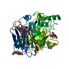
| Title | Crystal structure of intracellular family 1 beta-glucosidase BGL1A from the basidiomycete Phanerochaete chrysosporium in complex with gluconolactone | ||||||
 Components Components | Beta-glucosidase | ||||||
 Keywords Keywords | HYDROLASE / TIM BARREL / GLYCOSIDE HYDROLASE FAMILY 1 / CLAN GH-A / Structural Genomics / NPPSFA / National Project on Protein Structural and Functional Analyses | ||||||
| Function / homology |  Function and homology information Function and homology informationcellobiose glucosidase activity / beta-glucosidase / beta-glucosidase activity / cellulose catabolic process Similarity search - Function | ||||||
| Biological species |  Phanerochaete chrysosporium (fungus) Phanerochaete chrysosporium (fungus) | ||||||
| Method |  X-RAY DIFFRACTION / SYNCHROTRON / MOLECULAR REPLACEMENT / Resolution: 1.9 Å X-RAY DIFFRACTION / SYNCHROTRON / MOLECULAR REPLACEMENT / Resolution: 1.9 Å | ||||||
 Authors Authors | Nijikken, Y. / Tsukada, T. / Igarashi, K. / Samejima, M. / Fushinobu, S. | ||||||
 Citation Citation | Journal: Febs Lett. / Year: 2007 Title: Crystal structure of intracellular family 1 beta-glucosidase BGL1A from the basidiomycete Phanerochaete chrysosporium Authors: Nijikken, Y. / Tsukada, T. / Igarashi, K. / Samejima, M. / Wakagi, T. / Shoun, H. / Fushinobu, S. #1: Journal: Appl.Microbiol.Biotechnol. / Year: 2006 Title: Molecular cloning and characterization of two intracellular beta-glucosidases belonging to glycoside hydrolase family 1 from the basidiomycete Phanerochaete chrysosporium Authors: Tsukada, T. / Igarashi, K. / Yoshida, M. / Samejima, M. | ||||||
| History |
|
- Structure visualization
| Structure viewer | Molecule: MolmilJmol/JSmol |
|---|
- Downloads & links
-Download
| PDBx/mmCIF format | 2e40.cif.gz | 214.5 KB | Display | PDBx/mmCIF format |
|---|---|---|---|---|
| PDB format | pdb2e40.ent.gz | 168.4 KB | Display | PDB format |
| PDBx/mmJSON format | 2e40.json.gz | Tree view | PDBx/mmJSON format | |
| Others |  Other downloads Other downloads |
-Validation report
| Arichive directory | https://data.pdbj.org/pub/pdb/validation_reports/e4/2e40ftp://data.pdbj.org/pub/pdb/validation_reports/e4/2e40 | HTTPS FTP |
|---|
-Related structure data
| Related structure data |  2e3zC  1cbgS C: citing same article ( S: Starting model for refinement |
|---|---|
| Similar structure data |


-Links
 PDBj
PDBj- Assembly
| Deposited unit | 
| ||||||||
|---|---|---|---|---|---|---|---|---|---|
| 1 | 
| ||||||||
| 2 | 
| ||||||||
| Unit cell |
| ||||||||
| Details | The biological assembly is a monomer. The asymmetric unit contains two almost identical monomers. |
-Components
| #1: Protein | Mass: 52959.008 Da / Num. of mol.: 2 Source method: isolated from a genetically manipulated source Source: (gene. exp.) Phanerochaete chrysosporium (fungus) / Strain: K-3 / Gene: bgl1A / Plasmid: pBAD/TOPO ThioFusion / Production host:  #2: Sugar |   Type: D-saccharide / Mass: 178.140 Da / Num. of mol.: 2 Type: D-saccharide / Mass: 178.140 Da / Num. of mol.: 2Source method: isolated from a genetically manipulated source Formula: C6H10O6 #3: Water | ChemComp-HOH / |  Mass: 18.015 Da / Num. of mol.: 921 / Source method: isolated from a natural source / Formula: H2O Mass: 18.015 Da / Num. of mol.: 921 / Source method: isolated from a natural source / Formula: H2O |
|---|
-Experimental details
-Experiment
| Experiment | Method: X-RAY DIFFRACTION / Number of used crystals: 1 |
|---|
- Sample preparation
| Crystal | Density Matthews: 2.48 Å3/Da / Density % sol: 50.4 % |
|---|---|
| Crystal grow | Temperature: 298 K / Method: vapor diffusion, sitting drop / pH: 5.8 Details: 15% PEG 6000, 12% isopropanol, 0.1M sodium citrate, pH 5.8, VAPOR DIFFUSION, SITTING DROP, temperature 298K |
-Data collection
| Diffraction | Mean temperature: 100 K |
|---|---|
| Diffraction source | Source: SYNCHROTRON / Site: Photon Factory  / Beamline: BL-6A / Wavelength: 1 Å / Beamline: BL-6A / Wavelength: 1 Å |
| Detector | Type: ADSC QUANTUM 4 / Detector: CCD / Date: Dec 5, 2004 |
| Radiation | Monochromator: Triangular Si(111) with an asymmetric angle of 7.8 deg Protocol: SINGLE WAVELENGTH / Monochromatic (M) / Laue (L): M / Scattering type: x-ray |
| Radiation wavelength | Wavelength: 1 Å / Relative weight: 1 |
| Reflection | Resolution: 1.9→50 Å / Num. obs: 83722 / % possible obs: 99.5 % / Observed criterion σ(I): 0 / Redundancy: 4.2 % / Biso Wilson estimate: 15.5 Å2 / Rmerge(I) obs: 0.078 / Net I/σ(I): 19.4 |
| Reflection shell | Resolution: 1.9→1.97 Å / Rmerge(I) obs: 0.286 / Mean I/σ(I) obs: 3.59 / % possible all: 99.4 |
- Processing
| Software |
| ||||||||||||||||||||||||||||||||||||||||||||||||||||||||||||||||||||||||||||||||
|---|---|---|---|---|---|---|---|---|---|---|---|---|---|---|---|---|---|---|---|---|---|---|---|---|---|---|---|---|---|---|---|---|---|---|---|---|---|---|---|---|---|---|---|---|---|---|---|---|---|---|---|---|---|---|---|---|---|---|---|---|---|---|---|---|---|---|---|---|---|---|---|---|---|---|---|---|---|---|---|---|---|
| Refinement | Method to determine structure: MOLECULAR REPLACEMENT Starting model: PDB ENTRY 1CBG Resolution: 1.9→44.57 Å / Rfactor Rfree error: 0.003 / Data cutoff high absF: 2484620.95 / Data cutoff low absF: 0 / Isotropic thermal model: RESTRAINED / Cross valid method: THROUGHOUT / σ(F): 0 / Stereochemistry target values: Engh & Huber
| ||||||||||||||||||||||||||||||||||||||||||||||||||||||||||||||||||||||||||||||||
| Solvent computation | Solvent model: FLAT MODEL / Bsol: 45.7929 Å2 / ksol: 0.361751 e/Å3 | ||||||||||||||||||||||||||||||||||||||||||||||||||||||||||||||||||||||||||||||||
| Displacement parameters | Biso mean: 21.5 Å2
| ||||||||||||||||||||||||||||||||||||||||||||||||||||||||||||||||||||||||||||||||
| Refine analyze |
| ||||||||||||||||||||||||||||||||||||||||||||||||||||||||||||||||||||||||||||||||
| Refinement step | Cycle: LAST / Resolution: 1.9→44.57 Å
| ||||||||||||||||||||||||||||||||||||||||||||||||||||||||||||||||||||||||||||||||
| Refine LS restraints |
| ||||||||||||||||||||||||||||||||||||||||||||||||||||||||||||||||||||||||||||||||
| LS refinement shell | Resolution: 1.9→2.02 Å / Rfactor Rfree error: 0.009 / Total num. of bins used: 6
| ||||||||||||||||||||||||||||||||||||||||||||||||||||||||||||||||||||||||||||||||
| Xplor file |
|
