 Movie
Movie Controller

[English] 日本語
 Yorodumi
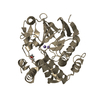
- PDB-2dyy: Crystal structure of putative translation initiation inhibitor PH... -
+ Open data

- Basic information
| Entry | Database: PDB / ID: 2dyy | ||||||
|---|---|---|---|---|---|---|---|
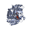
| Title | Crystal structure of putative translation initiation inhibitor PH0854 from Pyrococcus horikoshii | ||||||
 Components Components | UPF0076 protein PH0854 | ||||||
 Keywords Keywords | STRUCTURAL GENOMICS / UNKNOWN FUNCTION / Putative translation initiation inhibitor / Pyrococcus horikoshii / trimer / NPPSFA / National Project on Protein Structural and Functional Analyses / RIKEN Structural Genomics/Proteomics Initiative / RSGI | ||||||
| Function / homology |  Function and homology information Function and homology information | ||||||
| Biological species |   Pyrococcus horikoshii (archaea) Pyrococcus horikoshii (archaea) | ||||||
| Method |  X-RAY DIFFRACTION / SYNCHROTRON / MOLECULAR REPLACEMENT / Resolution: 2.6 Å X-RAY DIFFRACTION / SYNCHROTRON / MOLECULAR REPLACEMENT / Resolution: 2.6 Å | ||||||
 Authors Authors | Ihsanawati / Kishishita, S. / Murayama, K. / Chen, L. / Liu, Z.J. / Wang, B.C. / Shirouzu, M. / Bessho, Y. / Yokoyama, S. / RIKEN Structural Genomics/Proteomics Initiative (RSGI) | ||||||
 Citation Citation | Journal: To be Published Title: Crystal structure of putative translation initiation inhibitor PH0854 from Pyrococcus horikoshii Authors: Ihsanawati / Kishishita, S. / Murayama, K. / Chen, L. / Liu, Z.J. / Wang, B.C. / Shirouzu, M. / Bessho, Y. / Yokoyama, S. | ||||||
| History |
|
- Structure visualization
| Structure viewer | Molecule: MolmilJmol/JSmol |
|---|
- Downloads & links
-Download
| PDBx/mmCIF format | 2dyy.cif.gz | 282 KB | Display | PDBx/mmCIF format |
|---|---|---|---|---|
| PDB format | pdb2dyy.ent.gz | 231 KB | Display | PDB format |
| PDBx/mmJSON format | 2dyy.json.gz | Tree view | PDBx/mmJSON format | |
| Others |  Other downloads Other downloads |
-Validation report
| Summary document | 2dyy_validation.pdf.gz | 512.6 KB | Display | wwPDB validaton report |
|---|---|---|---|---|
| Full document | 2dyy_full_validation.pdf.gz | 550.2 KB | Display | |
| Data in XML | 2dyy_validation.xml.gz | 56.2 KB | Display | |
| Data in CIF | 2dyy_validation.cif.gz | 76.3 KB | Display | |
| Arichive directory | https://data.pdbj.org/pub/pdb/validation_reports/dy/2dyyftp://data.pdbj.org/pub/pdb/validation_reports/dy/2dyy | HTTPS FTP |
-Related structure data
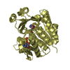
| Related structure data | 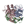 1xrgS S: Starting model for refinement |
|---|---|
| Similar structure data | |
| Other databases |


-Links
 PDBj
PDBj
- Assembly
| Deposited unit | 
| ||||||||
|---|---|---|---|---|---|---|---|---|---|
| 1 | 
| ||||||||
| 2 | 
| ||||||||
| 3 | 
| ||||||||
| 4 | 
| ||||||||
| Unit cell |
|
-Components
| #1: Protein | Mass: 14052.204 Da / Num. of mol.: 12 Source method: isolated from a genetically manipulated source Source: (gene. exp.) Pyrococcus horikoshii (archaea) / Plasmid: pET11a / Production host:  #2: Water | ChemComp-HOH / |  Mass: 18.015 Da / Num. of mol.: 233 / Source method: isolated from a natural source / Formula: H2O Mass: 18.015 Da / Num. of mol.: 233 / Source method: isolated from a natural source / Formula: H2O |
|---|
-Experimental details
-Experiment
| Experiment | Method: X-RAY DIFFRACTION / Number of used crystals: 1 |
|---|
- Sample preparation
| Crystal | Density Matthews: 2.01 Å3/Da / Density % sol: 38.8 % |
|---|---|
| Crystal grow | Temperature: 293 K / Method: vapor diffusion, hanging drop / pH: 7 Details: 2M NaCl, 0.1M TrisHCl pH 7.0, 30% (w/v) PEG 3000, VAPOR DIFFUSION, HANGING DROP, temperature 293K |
-Data collection
| Diffraction | Mean temperature: 100 K |
|---|---|
| Diffraction source | Source: SYNCHROTRON / Site: APS  / Beamline: 22-ID / Wavelength: 0.97914 Å / Beamline: 22-ID / Wavelength: 0.97914 Å |
| Detector | Type: MARMOSAIC 300 mm CCD / Detector: CCD / Date: Nov 26, 2005 |
| Radiation | Protocol: SINGLE WAVELENGTH / Monochromatic (M) / Laue (L): M / Scattering type: x-ray |
| Radiation wavelength | Wavelength: 0.97914 Å / Relative weight: 1 |
| Reflection | Resolution: 2.6→30 Å / Num. obs: 44546 / % possible obs: 96.6 % / Observed criterion σ(I): -3 / Biso Wilson estimate: 39.2 Å2 / Rmerge(I) obs: 0.096 |
| Reflection shell | Resolution: 2.6→2.76 Å / % possible all: 80.2 |
- Processing
| Software |
| ||||||||||||||||||||
|---|---|---|---|---|---|---|---|---|---|---|---|---|---|---|---|---|---|---|---|---|---|
| Refinement | Method to determine structure: MOLECULAR REPLACEMENT Starting model: 1XRG Resolution: 2.6→29.73 Å / Rfactor Rfree error: 0.007 / Data cutoff high absF: 1706561.8 / Data cutoff low absF: 0 / Isotropic thermal model: RESTRAINED / Cross valid method: THROUGHOUT / σ(F): 0
| ||||||||||||||||||||
| Solvent computation | Solvent model: FLAT MODEL / Bsol: 43.6057 Å2 / ksol: 0.35284 e/Å3 | ||||||||||||||||||||
| Displacement parameters | Biso mean: 42.1 Å2
| ||||||||||||||||||||
| Refine analyze |
| ||||||||||||||||||||
| Refinement step | Cycle: LAST / Resolution: 2.6→29.73 Å
| ||||||||||||||||||||
| Refine LS restraints |
| ||||||||||||||||||||
| LS refinement shell | Resolution: 2.6→2.76 Å / Rfactor Rfree error: 0.021 / Total num. of bins used: 6
| ||||||||||||||||||||
| Xplor file |
|
