 Movie
Movie Controller

[English] 日本語
 Yorodumi
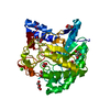
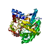
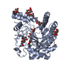
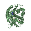
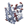
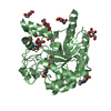
- PDB-2bvw: CELLOBIOHYDROLASE II (CEL6A) FROM HUMICOLA INSOLENS IN COMPLEX WI... -
+ Open data

- Basic information
| Entry | Database: PDB / ID: 2bvw | |||||||||
|---|---|---|---|---|---|---|---|---|---|---|
| Title | CELLOBIOHYDROLASE II (CEL6A) FROM HUMICOLA INSOLENS IN COMPLEX WITH GLUCOSE AND CELLOTETRAOSE | |||||||||
 Components Components | CELLOBIOHYDROLASE II | |||||||||
 Keywords Keywords | HYDROLASE / CELLULOSE DEGRADATION / CELLOBIOHYDROLASE / CELLULASE / GLYCOSIDE HYDROLASE FAMILY 6 | |||||||||
| Function / homology |  Function and homology information Function and homology informationcellulose 1,4-beta-cellobiosidase (non-reducing end) / cellulose 1,4-beta-cellobiosidase activity / cellulose binding / cellulose catabolic process / extracellular region Similarity search - Function | |||||||||
| Biological species |  Humicola insolens (fungus) Humicola insolens (fungus) | |||||||||
| Method |  X-RAY DIFFRACTION / SYNCHROTRON / MOLECULAR REPLACEMENT / Resolution: 1.7 Å X-RAY DIFFRACTION / SYNCHROTRON / MOLECULAR REPLACEMENT / Resolution: 1.7 Å | |||||||||
 Authors Authors | Varrot, A. / Davies, G.J. / Schulein, M. | |||||||||
 Citation Citation | Journal: Biochemistry / Year: 1999 Title: Structural changes of the active site tunnel of Humicola insolens cellobiohydrolase, Cel6A, upon oligosaccharide binding. Authors: Varrot, A. / Schulein, M. / Davies, G.J. | |||||||||
| History |
|
- Structure visualization
| Structure viewer | Molecule: MolmilJmol/JSmol |
|---|
- Downloads & links
-Download
| PDBx/mmCIF format | 2bvw.cif.gz | 170.9 KB | Display | PDBx/mmCIF format |
|---|---|---|---|---|
| PDB format | pdb2bvw.ent.gz | 133.9 KB | Display | PDB format |
| PDBx/mmJSON format | 2bvw.json.gz | Tree view | PDBx/mmJSON format | |
| Others |  Other downloads Other downloads |
-Validation report
| Arichive directory | https://data.pdbj.org/pub/pdb/validation_reports/bv/2bvwftp://data.pdbj.org/pub/pdb/validation_reports/bv/2bvw | HTTPS FTP |
|---|
-Related structure data
| Related structure data | 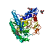 1bvwS S: Starting model for refinement |
|---|---|
| Similar structure data |


-Links
 PDBj
PDBj


- Assembly
| Deposited unit | 
| ||||||||
|---|---|---|---|---|---|---|---|---|---|
| 1 | 
| ||||||||
| 2 | 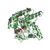
| ||||||||
| Unit cell |
| ||||||||
| Noncrystallographic symmetry (NCS) | NCS oper: (Code: given Matrix: (0.51751, -0.05441, 0.85394), Vector: |
-Components
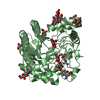
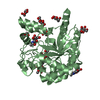
-Protein , 1 types, 2 molecules AB
| #1: Protein | Mass: 40014.512 Da / Num. of mol.: 2 / Fragment: CATALYTIC CORE DOMAIN Source method: isolated from a genetically manipulated source Details: N-LINKED N-ACETYLGLUCOSAMINE ON RESIDUE ASN 141 / Source: (gene. exp.) Humicola insolens (fungus) / Description: FULL-LENGTH ENZYME OVER-EXPRESSEDPlasmid: UNDER CONTROL OF THE FUNGAL AMYLASE PROMOTER AND AMYLOGLUCOSIDASE TERMINATOR Production host: References: UniProt: Q9C1S9, cellulose 1,4-beta-cellobiosidase (non-reducing end) |
|---|
-Sugars , 4 types, 6 molecules 

| #2: Polysaccharide | beta-D-glucopyranose-(1-4)-beta-D-glucopyranose-(1-4)-beta-D-glucopyranose-(1-4)-beta-D-glucopyranose / beta-cellotetraose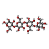  Source method: isolated from a genetically manipulated source Details: oligosaccharide / References: beta-cellotetraose | ||
|---|---|---|---|
| #3: Polysaccharide | beta-D-glucopyranose-(1-4)-beta-D-glucopyranose-(1-4)-alpha-D-glucopyranose / alpha-cellotriose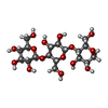  Source method: isolated from a genetically manipulated source Details: oligosaccharide / References: alpha-cellotriose | ||
| #4: Sugar |  Type: D-saccharide, beta linking / Mass: 221.208 Da / Num. of mol.: 2 Type: D-saccharide, beta linking / Mass: 221.208 Da / Num. of mol.: 2Source method: isolated from a genetically manipulated source Formula: C8H15NO6 #5: Sugar |  Type: D-saccharide, beta linking / Mass: 180.156 Da / Num. of mol.: 2 / Source method: obtained synthetically / Formula: C6H12O6 Type: D-saccharide, beta linking / Mass: 180.156 Da / Num. of mol.: 2 / Source method: obtained synthetically / Formula: C6H12O6 |
-Non-polymers , 2 types, 624 molecules 

| #6: Chemical | ChemComp-GOL /  Mass: 92.094 Da / Num. of mol.: 7 / Source method: obtained synthetically / Formula: C3H8O3 Mass: 92.094 Da / Num. of mol.: 7 / Source method: obtained synthetically / Formula: C3H8O3#7: Water | ChemComp-HOH / | Mass: 18.015 Da / Num. of mol.: 617 / Source method: isolated from a natural source / Formula: H2O |
|---|
-Details
| Has protein modification | Y |
|---|---|
| Nonpolymer details | CELLOTETRAOSE WITH ALPHA AND BETA OXYGENE AT C1 CELLOTRIOSE WITH ALPHA AND BETA OXYGENE AT C1 ...CELLOTETRA |
| Sequence details | THERE IS A PRO-SEQUENCE OF 23 AMINO-ACIDS WHICH ARE POST-TRANSLATIONALLY CLEAVED TO YIELD A MATURE ...THERE IS A PRO-SEQUENCE OF 23 AMINO-ACIDS WHICH ARE POST-TRANSLATIO |
-Experimental details
-Experiment
| Experiment | Method: X-RAY DIFFRACTION / Number of used crystals: 2 |
|---|
- Sample preparation
| Crystal | Density Matthews: 2.01 Å3/Da / Density % sol: 38.2 % Description: SEARCH MODEL WAS CEL6A FROM HUMICOLA INSOLENS NATIVE STRUCTURE | ||||||||||||||||||||||||||||||||||||
|---|---|---|---|---|---|---|---|---|---|---|---|---|---|---|---|---|---|---|---|---|---|---|---|---|---|---|---|---|---|---|---|---|---|---|---|---|---|
| Crystal grow | pH: 4.6 / Details: pH 4.6 | ||||||||||||||||||||||||||||||||||||
| Crystal grow | *PLUS Method: vapor diffusion, hanging dropDetails: drop contains a 50:50(v/v) mix of protein and of reservoir solution | ||||||||||||||||||||||||||||||||||||
| Components of the solutions | *PLUS
|
-Data collection
| Diffraction | Mean temperature: 100 K |
|---|---|
| Diffraction source | Source: SYNCHROTRON / Site: SRS  / Beamline: PX9.6 / Wavelength: 0.87 / Beamline: PX9.6 / Wavelength: 0.87 |
| Detector | Type: ADSC QUANTUM 4 / Detector: CCD / Date: Jul 1, 1998 / Details: TORROIDAL MIRROR |
| Radiation | Monochromator: SILICON / Protocol: SINGLE WAVELENGTH / Monochromatic (M) / Laue (L): M / Scattering type: x-ray |
| Radiation wavelength | Wavelength: 0.87 Å / Relative weight: 1 |
| Reflection | Resolution: 1.7→20 Å / Num. obs: 72987 / % possible obs: 89.6 % / Redundancy: 4.4 % / Rmerge(I) obs: 0.078 / Rsym value: 0.078 / Net I/σ(I): 11.4 |
| Reflection shell | Resolution: 1.7→1.79 Å / Redundancy: 4.6 % / Rmerge(I) obs: 0.359 / Mean I/σ(I) obs: 3.9 / Rsym value: 0.359 / % possible all: 89.6 |
| Reflection shell | *PLUS % possible obs: 89.6 % / Mean I/σ(I) obs: 1.9 |
- Processing
| Software |
| ||||||||||||||||||||||||||||||||||||||||||||||||||||||||||||||||||||||||||||||||||||
|---|---|---|---|---|---|---|---|---|---|---|---|---|---|---|---|---|---|---|---|---|---|---|---|---|---|---|---|---|---|---|---|---|---|---|---|---|---|---|---|---|---|---|---|---|---|---|---|---|---|---|---|---|---|---|---|---|---|---|---|---|---|---|---|---|---|---|---|---|---|---|---|---|---|---|---|---|---|---|---|---|---|---|---|---|---|
| Refinement | Method to determine structure: MOLECULAR REPLACEMENT Starting model: PDB ENTRY 1BVW Resolution: 1.7→20 Å / Cross valid method: THROUGHOUT / σ(F): 0
| ||||||||||||||||||||||||||||||||||||||||||||||||||||||||||||||||||||||||||||||||||||
| Refinement step | Cycle: LAST / Resolution: 1.7→20 Å
| ||||||||||||||||||||||||||||||||||||||||||||||||||||||||||||||||||||||||||||||||||||
| Refine LS restraints |
| ||||||||||||||||||||||||||||||||||||||||||||||||||||||||||||||||||||||||||||||||||||
| Software | *PLUS Name: CCP4 / Classification: refinement | ||||||||||||||||||||||||||||||||||||||||||||||||||||||||||||||||||||||||||||||||||||
| Refinement | *PLUS Rfactor Rfree: 0.225 | ||||||||||||||||||||||||||||||||||||||||||||||||||||||||||||||||||||||||||||||||||||
| Solvent computation | *PLUS | ||||||||||||||||||||||||||||||||||||||||||||||||||||||||||||||||||||||||||||||||||||
| Displacement parameters | *PLUS |
