 Movie
Movie Controller


+ Open data

- Basic information
| Entry | Database: PDB / ID: 1bvw | ||||||
|---|---|---|---|---|---|---|---|
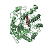
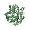
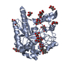
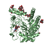
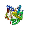
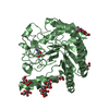
| Title | CELLOBIOHYDROLASE II (CEL6A) FROM HUMICOLA INSOLENS | ||||||
 Components Components | PROTEIN (CELLOBIOHYDROLASE II) | ||||||
 Keywords Keywords | HYDROLASE / CELLULOSE DEGRADATION / CELLOBIOHYDROLASE / CELLULASE / GLYCOSIDE HYDROLASE FAMILY 6 | ||||||
| Function / homology |  Function and homology information Function and homology informationcellulose 1,4-beta-cellobiosidase (non-reducing end) / cellulose 1,4-beta-cellobiosidase activity / cellulose binding / cellulose catabolic process / extracellular region Similarity search - Function | ||||||
| Biological species |  Humicola insolens (fungus) Humicola insolens (fungus) | ||||||
| Method |  X-RAY DIFFRACTION / SYNCHROTRON / MOLECULAR REPLACEMENT / Resolution: 1.92 Å X-RAY DIFFRACTION / SYNCHROTRON / MOLECULAR REPLACEMENT / Resolution: 1.92 Å | ||||||
 Authors Authors | Varrot, A. / Davies, G.J. / Schulein, M. | ||||||
 Citation Citation | Journal: Biochem.J. / Year: 1999 Title: Crystal structure of the catalytic core domain of the family 6 cellobiohydrolase II, Cel6A, from Humicola insolens, at 1.92 A resolution. Authors: Varrot, A. / Hastrup, S. / Schulein, M. / Davies, G.J. | ||||||
| History |
|
- Structure visualization
| Structure viewer | Molecule: MolmilJmol/JSmol |
|---|
- Downloads & links
-Download
| PDBx/mmCIF format | 1bvw.cif.gz | 98.8 KB | Display | PDBx/mmCIF format |
|---|---|---|---|---|
| PDB format | pdb1bvw.ent.gz | 72.1 KB | Display | PDB format |
| PDBx/mmJSON format | 1bvw.json.gz | Tree view | PDBx/mmJSON format | |
| Others |  Other downloads Other downloads |
-Validation report
| Arichive directory | https://data.pdbj.org/pub/pdb/validation_reports/bv/1bvwftp://data.pdbj.org/pub/pdb/validation_reports/bv/1bvw | HTTPS FTP |
|---|
-Related structure data
| Related structure data | 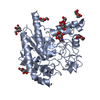 1cb2S S: Starting model for refinement |
|---|---|
| Similar structure data |


-Links
 PDBj
PDBj


- Assembly
| Deposited unit | 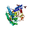
| ||||||||
|---|---|---|---|---|---|---|---|---|---|
| 1 |
| ||||||||
| Unit cell |
|
-Components
-Protein , 1 types, 1 molecules A
| #1: Protein | Mass: 39737.234 Da / Num. of mol.: 1 / Fragment: CATALYTIC CORE DOMAIN RESIDUES 91-450 Source method: isolated from a genetically manipulated source Details: N-LINKED N-ACETYLGLUCOSAMINE ON RESIDUE ASN 141 O-LINKED MANNOSE (ALPHA-LINKED) ON RESIDUE THR 118 O-LINKED MANNOSE (ALPHA-LINKED) ON RESIDUE SER 127 Source: (gene. exp.) Humicola insolens (fungus)Description: UNDER CONTROL OF THE FUNGAL AMYLASE PROMOTER AND AMYLOGLUCOSIDASE TERMINATOR Production host: References: UniProt: Q9C1S9, cellulose 1,4-beta-cellobiosidase (non-reducing end) |
|---|
-Sugars , 2 types, 3 molecules 

| #2: Sugar |  Type: D-saccharide, alpha linking / Mass: 180.156 Da / Num. of mol.: 2 Type: D-saccharide, alpha linking / Mass: 180.156 Da / Num. of mol.: 2Source method: isolated from a genetically manipulated source Formula: C6H12O6 #3: Sugar | ChemComp-NAG / |  Type: D-saccharide, beta linking / Mass: 221.208 Da / Num. of mol.: 1 Type: D-saccharide, beta linking / Mass: 221.208 Da / Num. of mol.: 1Source method: isolated from a genetically manipulated source Formula: C8H15NO6 |
|---|
-Non-polymers , 3 types, 533 molecules 


| #4: Chemical | ChemComp-MG /  Mass: 24.305 Da / Num. of mol.: 1 / Source method: obtained synthetically / Formula: Mg Mass: 24.305 Da / Num. of mol.: 1 / Source method: obtained synthetically / Formula: Mg |
|---|---|
| #5: Chemical | ChemComp-GOL /  Mass: 92.094 Da / Num. of mol.: 1 / Source method: obtained synthetically / Formula: C3H8O3 Mass: 92.094 Da / Num. of mol.: 1 / Source method: obtained synthetically / Formula: C3H8O3 |
| #6: Water | ChemComp-HOH / Mass: 18.015 Da / Num. of mol.: 531 / Source method: isolated from a natural source / Formula: H2O |
-Details
| Has protein modification | Y |
|---|
-Experimental details
-Experiment
| Experiment | Method: X-RAY DIFFRACTION / Number of used crystals: 1 |
|---|
- Sample preparation
| Crystal | Density Matthews: 2.01 Å3/Da / Density % sol: 42 % Description: SEARCH MODEL WAS THE Y169F MUTANT OF THE TRICHODERMA REESEI CELLOBIOHYDROLASE II | |||||||||||||||||||||||||
|---|---|---|---|---|---|---|---|---|---|---|---|---|---|---|---|---|---|---|---|---|---|---|---|---|---|---|
| Crystal grow | pH: 7 Details: PROTEIN WAS CONCENTRATED TO 20 MG/ML IN WATER. CRYSTALLISATION IN 200MM MAGNESIUM ACETATE IN 100MM TRIETHANOLAMINE BUFFER AT PH 7.0. PRECIPITANT WAS 22%. POLYETHYLENE GLYCOL 8000. | |||||||||||||||||||||||||
| Crystal | *PLUS | |||||||||||||||||||||||||
| Crystal grow | *PLUS pH: 7 / Method: vapor diffusion, hanging drop | |||||||||||||||||||||||||
| Components of the solutions | *PLUS
|
-Data collection
| Diffraction | Mean temperature: 100 K |
|---|---|
| Diffraction source | Source: SYNCHROTRON / Site: SRS  / Beamline: PX9.6 / Wavelength: 0.872 / Beamline: PX9.6 / Wavelength: 0.872 |
| Detector | Type: MARRESEARCH / Detector: IMAGE PLATE / Date: Dec 15, 1997 / Details: TORROIDAL MIRROR |
| Radiation | Monochromator: SILICON / Protocol: SINGLE WAVELENGTH / Monochromatic (M) / Laue (L): M / Scattering type: x-ray |
| Radiation wavelength | Wavelength: 0.872 Å / Relative weight: 1 |
| Reflection | Resolution: 1.92→20 Å / Num. obs: 90583 / % possible obs: 99.7 % / Observed criterion σ(I): 0 / Redundancy: 3.8 % / Biso Wilson estimate: 13.5 Å2 / Rmerge(I) obs: 0.063 / Rsym value: 0.063 / Net I/σ(I): 20 |
| Reflection shell | Resolution: 1.92→1.99 Å / Redundancy: 3.83 % / Rmerge(I) obs: 0.187 / Mean I/σ(I) obs: 7.3 / Rsym value: 0.187 / % possible all: 99.1 |
| Reflection shell | *PLUS % possible obs: 99.1 % |
- Processing
| Software |
| ||||||||||||||||||||||||||||||||||||||||||||||||||||||||||||||||||||||||||||||||||||
|---|---|---|---|---|---|---|---|---|---|---|---|---|---|---|---|---|---|---|---|---|---|---|---|---|---|---|---|---|---|---|---|---|---|---|---|---|---|---|---|---|---|---|---|---|---|---|---|---|---|---|---|---|---|---|---|---|---|---|---|---|---|---|---|---|---|---|---|---|---|---|---|---|---|---|---|---|---|---|---|---|---|---|---|---|---|
| Refinement | Method to determine structure: MOLECULAR REPLACEMENT Starting model: PDB ENTRY 1CB2 Resolution: 1.92→15 Å / Cross valid method: THROUGHOUT / σ(F): 0
| ||||||||||||||||||||||||||||||||||||||||||||||||||||||||||||||||||||||||||||||||||||
| Displacement parameters | Biso mean: 14.3 Å2 | ||||||||||||||||||||||||||||||||||||||||||||||||||||||||||||||||||||||||||||||||||||
| Refinement step | Cycle: LAST / Resolution: 1.92→15 Å
| ||||||||||||||||||||||||||||||||||||||||||||||||||||||||||||||||||||||||||||||||||||
| Refine LS restraints |
| ||||||||||||||||||||||||||||||||||||||||||||||||||||||||||||||||||||||||||||||||||||
| Software | *PLUS Name: REFMAC / Classification: refinement | ||||||||||||||||||||||||||||||||||||||||||||||||||||||||||||||||||||||||||||||||||||
| Refinement | *PLUS Lowest resolution: 15 Å / σ(F): 0 / % reflection Rfree: 5 % / Rfactor obs: 0.14 / Rfactor Rfree: 0.21 | ||||||||||||||||||||||||||||||||||||||||||||||||||||||||||||||||||||||||||||||||||||
| Solvent computation | *PLUS | ||||||||||||||||||||||||||||||||||||||||||||||||||||||||||||||||||||||||||||||||||||
| Displacement parameters | *PLUS Biso mean: 14.3 Å2 | ||||||||||||||||||||||||||||||||||||||||||||||||||||||||||||||||||||||||||||||||||||
| Refine LS restraints | *PLUS Type: p_plane_restr / Dev ideal: 0.019 / Dev ideal target: 0.03 |
