 Movie
Movie Controller


+ Open data

- Basic information
| Entry | Database: PDB / ID: 1cb2 | ||||||
|---|---|---|---|---|---|---|---|
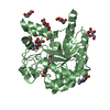
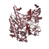
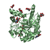
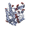
| Title | CELLOBIOHYDROLASE II, CATALYTIC DOMAIN, MUTANT Y169F | ||||||
 Components Components | CELLOBIOHYDROLASE II | ||||||
 Keywords Keywords | HYDROLASE (O-GLYCOSYL) / GLYCOSIDASE / GLYCOPROTEIN | ||||||
| Function / homology |  Function and homology information Function and homology informationcellulose 1,4-beta-cellobiosidase (non-reducing end) / cellulose 1,4-beta-cellobiosidase activity / cellulose binding / cellulose catabolic process / extracellular region / identical protein binding Similarity search - Function | ||||||
| Biological species |  Hypocrea jecorina (fungus) Hypocrea jecorina (fungus) | ||||||
| Method |  X-RAY DIFFRACTION / Resolution: 2 Å X-RAY DIFFRACTION / Resolution: 2 Å | ||||||
 Authors Authors | Kleywegt, G.J. / Szardenings, M. / Jones, T.A. | ||||||
 Citation Citation | Journal: Protein Eng. / Year: 1996 Title: The active site of Trichoderma reesei cellobiohydrolase II: the role of tyrosine 169. Authors: Koivula, A. / Reinikainen, T. / Ruohonen, L. / Valkeajarvi, A. / Claeyssens, M. / Teleman, O. / Kleywegt, G.J. / Szardenings, M. / Rouvinen, J. / Jones, T.A. / Teeri, T.T. #1: Journal: Science / Year: 1990Title: Three-Dimensional Structure of Cellobiohydrolase II from Trichoderma Reesei Authors: Rouvinen, J. / Bergfors, T. / Teeri, T. / Knowles, J.K. / Jones, T.A. | ||||||
| History |
|
- Structure visualization
| Structure viewer | Molecule: MolmilJmol/JSmol |
|---|
- Downloads & links
-Download
| PDBx/mmCIF format | 1cb2.cif.gz | 146.9 KB | Display | PDBx/mmCIF format |
|---|---|---|---|---|
| PDB format | pdb1cb2.ent.gz | 117.4 KB | Display | PDB format |
| PDBx/mmJSON format | 1cb2.json.gz | Tree view | PDBx/mmJSON format | |
| Others |  Other downloads Other downloads |
-Validation report
| Arichive directory | https://data.pdbj.org/pub/pdb/validation_reports/cb/1cb2ftp://data.pdbj.org/pub/pdb/validation_reports/cb/1cb2 | HTTPS FTP |
|---|
-Related structure data
| Similar structure data |
|---|


-Links
 PDBj
PDBj


- Assembly
| Deposited unit | 
| ||||||||
|---|---|---|---|---|---|---|---|---|---|
| 1 | 
| ||||||||
| 2 | 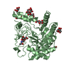
| ||||||||
| Unit cell |
| ||||||||
| Noncrystallographic symmetry (NCS) | NCS oper: (Code: given Matrix: (0.994635, -0.096273, -0.037846), Vector: |
-Components
| #1: Protein | Mass: 39039.297 Da / Num. of mol.: 2 / Fragment: CATALYTIC / Mutation: Y169F Source method: isolated from a genetically manipulated source Source: (gene. exp.) Hypocrea jecorina (fungus) / Gene: CBH2 (Y169F) / Gene (production host): CBH2 (Y169F) / Production host: Hypocrea jecorina (fungus)References: UniProt: P07987, cellulose 1,4-beta-cellobiosidase (non-reducing end) #2: Sugar | ChemComp-NAG /   Type: D-saccharide, beta linking / Mass: 221.208 Da / Num. of mol.: 4 Type: D-saccharide, beta linking / Mass: 221.208 Da / Num. of mol.: 4Source method: isolated from a genetically manipulated source Formula: C8H15NO6 #3: Sugar | ChemComp-MAN /   Type: D-saccharide, alpha linking / Mass: 180.156 Da / Num. of mol.: 14 Type: D-saccharide, alpha linking / Mass: 180.156 Da / Num. of mol.: 14Source method: isolated from a genetically manipulated source Formula: C6H12O6 #4: Water | ChemComp-HOH / |  Mass: 18.015 Da / Num. of mol.: 392 / Source method: isolated from a natural source / Formula: H2O Mass: 18.015 Da / Num. of mol.: 392 / Source method: isolated from a natural source / Formula: H2OCompound details | THE CATALYTIC CORE STARTS AT RESIDUE 83. | Has protein modification | Y | |
|---|
-Experimental details
-Experiment
| Experiment | Method: X-RAY DIFFRACTION |
|---|
- Sample preparation
| Crystal | Density Matthews: 2.15 Å3/Da / Density % sol: 42.91 % | ||||||||||||||||||||||||||||||
|---|---|---|---|---|---|---|---|---|---|---|---|---|---|---|---|---|---|---|---|---|---|---|---|---|---|---|---|---|---|---|---|
| Crystal grow | *PLUS pH: 6 / Method: vapor diffusion, hanging drop | ||||||||||||||||||||||||||||||
| Components of the solutions | *PLUS
|
-Data collection
| Diffraction source | Wavelength: 1.5418 |
|---|---|
| Detector | Type: XUONG-HAMLIN MULTIWIRE / Detector: AREA DETECTOR / Date: Apr 2, 1993 |
| Radiation | Monochromatic (M) / Laue (L): M / Scattering type: x-ray |
| Radiation wavelength | Wavelength: 1.5418 Å / Relative weight: 1 |
| Reflection | Num. obs: 46849 / % possible obs: 91.7 % / Redundancy: 4.3 % / Rmerge(I) obs: 0.068 |
| Reflection | *PLUS Highest resolution: 1.8 Å / Num. measured all: 202105 |
- Processing
| Software |
| ||||||||||||||||||||||||||||||||||||||||||||||||||||||||||||
|---|---|---|---|---|---|---|---|---|---|---|---|---|---|---|---|---|---|---|---|---|---|---|---|---|---|---|---|---|---|---|---|---|---|---|---|---|---|---|---|---|---|---|---|---|---|---|---|---|---|---|---|---|---|---|---|---|---|---|---|---|---|
| Refinement | Resolution: 2→8 Å / σ(F): 0
| ||||||||||||||||||||||||||||||||||||||||||||||||||||||||||||
| Displacement parameters | Biso mean: 15.8 Å2 | ||||||||||||||||||||||||||||||||||||||||||||||||||||||||||||
| Refinement step | Cycle: LAST / Resolution: 2→8 Å
| ||||||||||||||||||||||||||||||||||||||||||||||||||||||||||||
| Refine LS restraints |
| ||||||||||||||||||||||||||||||||||||||||||||||||||||||||||||
| Software | *PLUS Name: X-PLOR / Classification: refinement | ||||||||||||||||||||||||||||||||||||||||||||||||||||||||||||
| Refinement | *PLUS | ||||||||||||||||||||||||||||||||||||||||||||||||||||||||||||
| Solvent computation | *PLUS | ||||||||||||||||||||||||||||||||||||||||||||||||||||||||||||
| Displacement parameters | *PLUS | ||||||||||||||||||||||||||||||||||||||||||||||||||||||||||||
| Refine LS restraints | *PLUS
|
