 Movie
Movie Controller

[English] 日本語
 Yorodumi
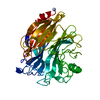
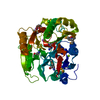
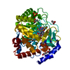
- PDB-1rje: Structure of PPM1, a leucine carboxy methyltransferase involved i... -
+ Open data

- Basic information
| Entry | Database: PDB / ID: 1rje | ||||||
|---|---|---|---|---|---|---|---|
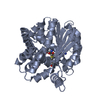
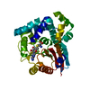
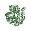
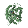
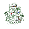
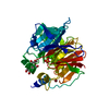
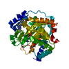
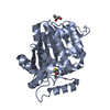
| Title | Structure of PPM1, a leucine carboxy methyltransferase involved in the regulation of protein phosphatase 2A activity | ||||||
 Components Components | carboxy methyl transferase for protein phosphatase 2A catalytic subunit | ||||||
 Keywords Keywords | TRANSFERASE / SAM dependent methyltransferase | ||||||
| Function / homology |  Function and homology information Function and homology informationCyclin A/B1/B2 associated events during G2/M transition / [phosphatase 2A protein]-leucine-carboxy methyltransferase / protein C-terminal leucine carboxyl O-methyltransferase activity / regulation of autophagy / protein-containing complex assembly / methylation Similarity search - Function | ||||||
| Biological species |  | ||||||
| Method |  X-RAY DIFFRACTION / SYNCHROTRON / MOLECULAR REPLACEMENT / Resolution: 2 Å X-RAY DIFFRACTION / SYNCHROTRON / MOLECULAR REPLACEMENT / Resolution: 2 Å | ||||||
 Authors Authors | Leulliot, N. / Quevillon-Cheruel, S. / Sorel, I. / de La Sierra-Gallay, I.L. / Collinet, B. / Graille, M. / Blondeau, K. / Bettache, N. / Poupon, A. / Janin, J. / van Tilbeurgh, H. | ||||||
 Citation Citation | Journal: J.Biol.Chem. / Year: 2004 Title: Structure of protein phosphatase methyltransferase 1 (PPM1), a leucine carboxyl methyltransferase involved in the regulation of protein phosphatase 2A activity. Authors: Leulliot, N. / Quevillon-Cheruel, S. / Sorel, I. / de La Sierra-Gallay, I.L. / Collinet, B. / Graille, M. / Blondeau, K. / Bettache, N. / Poupon, A. / Janin, J. / van Tilbeurgh, H. | ||||||
| History |
|
- Structure visualization
| Structure viewer | Molecule: MolmilJmol/JSmol |
|---|
- Downloads & links
-Download
| PDBx/mmCIF format | 1rje.cif.gz | 233.6 KB | Display | PDBx/mmCIF format |
|---|---|---|---|---|
| PDB format | pdb1rje.ent.gz | 186 KB | Display | PDB format |
| PDBx/mmJSON format | 1rje.json.gz | Tree view | PDBx/mmJSON format | |
| Others |  Other downloads Other downloads |
-Validation report
| Arichive directory | https://data.pdbj.org/pub/pdb/validation_reports/rj/1rjeftp://data.pdbj.org/pub/pdb/validation_reports/rj/1rje | HTTPS FTP |
|---|
-Related structure data


-Links
 PDBj
PDBj


- Assembly
| Deposited unit | 
| ||||||||
|---|---|---|---|---|---|---|---|---|---|
| 1 | 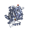
| ||||||||
| 2 | 
| ||||||||
| 3 | 
| ||||||||
| Unit cell |
|
-Components
| #1: Protein | Mass: 38567.438 Da / Num. of mol.: 3 Source method: isolated from a genetically manipulated source Source: (gene. exp.) Gene: PPM1 / Plasmid: pET9 / Production host:  References: UniProt: Q04081, Transferases; Transferring one-carbon groups; Methyltransferases #2: Chemical |   Mass: 96.063 Da / Num. of mol.: 2 / Source method: obtained synthetically / Formula: SO4 Mass: 96.063 Da / Num. of mol.: 2 / Source method: obtained synthetically / Formula: SO4#3: Chemical |   Type: L-peptide linking / Mass: 384.411 Da / Num. of mol.: 3 / Source method: obtained synthetically / Formula: C14H20N6O5S Type: L-peptide linking / Mass: 384.411 Da / Num. of mol.: 3 / Source method: obtained synthetically / Formula: C14H20N6O5S#4: Chemical | ChemComp-BME /   Mass: 78.133 Da / Num. of mol.: 6 / Source method: obtained synthetically / Formula: C2H6OS Mass: 78.133 Da / Num. of mol.: 6 / Source method: obtained synthetically / Formula: C2H6OS#5: Water | ChemComp-HOH / |  Mass: 18.015 Da / Num. of mol.: 990 / Source method: isolated from a natural source / Formula: H2O Mass: 18.015 Da / Num. of mol.: 990 / Source method: isolated from a natural source / Formula: H2O |
|---|
-Experimental details
-Experiment
| Experiment | Method: X-RAY DIFFRACTION / Number of used crystals: 1 |
|---|
- Sample preparation
| Crystal | Density Matthews: 2.53 Å3/Da / Density % sol: 51.46 % | ||||||||||||||||||||||||
|---|---|---|---|---|---|---|---|---|---|---|---|---|---|---|---|---|---|---|---|---|---|---|---|---|---|
| Crystal grow | Temperature: 293 K / Method: vapor diffusion, hanging drop / pH: 5.6 Details: 15% PEG 8000, 0.2M ammonium sulfate, 0.1M MES, pH 5.6, VAPOR DIFFUSION, HANGING DROP, temperature 293K | ||||||||||||||||||||||||
| Crystal grow | *PLUS Temperature: 293 K / pH: 4.6 / Method: vapor diffusion, hanging drop | ||||||||||||||||||||||||
| Components of the solutions | *PLUS
|
-Data collection
| Diffraction | Mean temperature: 100 K |
|---|---|
| Diffraction source | Source: SYNCHROTRON / Site: ESRF  / Beamline: ID14-4 / Wavelength: 0.933 Å / Beamline: ID14-4 / Wavelength: 0.933 Å |
| Detector | Type: ADSC QUANTUM 4 / Detector: CCD / Date: Feb 14, 2002 |
| Radiation | Monochromator: Si111 / Protocol: SINGLE WAVELENGTH / Monochromatic (M) / Laue (L): M / Scattering type: x-ray |
| Radiation wavelength | Wavelength: 0.933 Å / Relative weight: 1 |
| Reflection | Resolution: 2→24 Å / Num. obs: 76511 / % possible obs: 98.8 % / Observed criterion σ(F): 2 / Observed criterion σ(I): 2 |
| Reflection shell | Resolution: 2→2.11 Å / % possible all: 92 |
| Reflection | *PLUS Highest resolution: 2 Å / Lowest resolution: 24 Å / Redundancy: 3.8 % / Num. measured all: 294159 / Rmerge(I) obs: 0.12 |
- Processing
| Software |
| ||||||||||||||||||||||||||||||||||||||||||||||||||||||||||||||||||||||||||||||||||||||||||||||||||||
|---|---|---|---|---|---|---|---|---|---|---|---|---|---|---|---|---|---|---|---|---|---|---|---|---|---|---|---|---|---|---|---|---|---|---|---|---|---|---|---|---|---|---|---|---|---|---|---|---|---|---|---|---|---|---|---|---|---|---|---|---|---|---|---|---|---|---|---|---|---|---|---|---|---|---|---|---|---|---|---|---|---|---|---|---|---|---|---|---|---|---|---|---|---|---|---|---|---|---|---|---|---|
| Refinement | Method to determine structure: MOLECULAR REPLACEMENT / Resolution: 2→24.47 Å / Cor.coef. Fo:Fc: 0.951 / Cor.coef. Fo:Fc free: 0.918 / SU B: 3.885 / SU ML: 0.107 / Cross valid method: THROUGHOUT / σ(F): 2 / ESU R: 0.163 / ESU R Free: 0.152 / Stereochemistry target values: MAXIMUM LIKELIHOOD / Details: HYDROGENS HAVE BEEN ADDED IN THE RIDING POSITIONS
| ||||||||||||||||||||||||||||||||||||||||||||||||||||||||||||||||||||||||||||||||||||||||||||||||||||
| Solvent computation | Ion probe radii: 0.8 Å / Shrinkage radii: 0.8 Å / VDW probe radii: 1.4 Å / Solvent model: BABINET MODEL WITH MASK | ||||||||||||||||||||||||||||||||||||||||||||||||||||||||||||||||||||||||||||||||||||||||||||||||||||
| Displacement parameters | Biso mean: 16.526 Å2
| ||||||||||||||||||||||||||||||||||||||||||||||||||||||||||||||||||||||||||||||||||||||||||||||||||||
| Refinement step | Cycle: LAST / Resolution: 2→24.47 Å
| ||||||||||||||||||||||||||||||||||||||||||||||||||||||||||||||||||||||||||||||||||||||||||||||||||||
| Refine LS restraints |
| ||||||||||||||||||||||||||||||||||||||||||||||||||||||||||||||||||||||||||||||||||||||||||||||||||||
| LS refinement shell | Resolution: 2→2.052 Å / Total num. of bins used: 20 /
| ||||||||||||||||||||||||||||||||||||||||||||||||||||||||||||||||||||||||||||||||||||||||||||||||||||
| Refinement | *PLUS Highest resolution: 2 Å / Lowest resolution: 24 Å / % reflection Rfree: 5 % / Rfactor Rfree: 0.21 / Rfactor Rwork: 0.153 | ||||||||||||||||||||||||||||||||||||||||||||||||||||||||||||||||||||||||||||||||||||||||||||||||||||
| Solvent computation | *PLUS | ||||||||||||||||||||||||||||||||||||||||||||||||||||||||||||||||||||||||||||||||||||||||||||||||||||
| Displacement parameters | *PLUS | ||||||||||||||||||||||||||||||||||||||||||||||||||||||||||||||||||||||||||||||||||||||||||||||||||||
| Refine LS restraints | *PLUS
|
