 Movie
Movie Controller


+ Open data

- Basic information
| Entry | Database: PDB / ID: 7k2u | ||||||
|---|---|---|---|---|---|---|---|
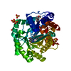
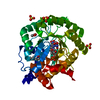
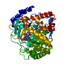
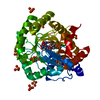
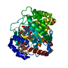
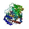
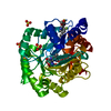
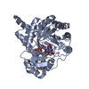
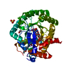
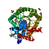
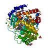
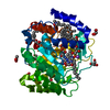
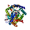
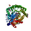
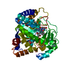
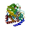
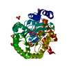
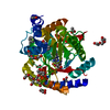
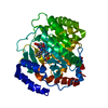
| Title | DHODH IN COMPLEX WITH LIGAND 13 | ||||||
 Components Components | Dihydroorotate dehydrogenase (quinone), mitochondrial | ||||||
 Keywords Keywords | OXIDOREDUCTASE / DIHYDROOROTATE DEHYDROGENASE / DHODH / INHIBITOR | ||||||
| Function / homology |  Function and homology information Function and homology informationpyrimidine ribonucleotide biosynthetic process / dihydroorotate dehydrogenase (quinone) / dihydroorotate dehydrogenase (quinone) activity / dihydroorotate dehydrogenase activity / dihydroorotase activity / Pyrimidine biosynthesis / UDP biosynthetic process / 'de novo' UMP biosynthetic process / 'de novo' pyrimidine nucleobase biosynthetic process / mitochondrial inner membrane / mitochondrion Similarity search - Function | ||||||
| Biological species |  Homo sapiens (human) Homo sapiens (human) | ||||||
| Method |  X-RAY DIFFRACTION / SYNCHROTRON / MOLECULAR REPLACEMENT / Resolution: 1.73 Å X-RAY DIFFRACTION / SYNCHROTRON / MOLECULAR REPLACEMENT / Resolution: 1.73 Å | ||||||
 Authors Authors | Shaffer, P.L. | ||||||
 Citation Citation | Journal: Bioorg.Med.Chem.Lett. / Year: 2020 Title: A carboxylic acid isostere screen of the DHODH inhibitor Brequinar. Authors: DeRatt, L.G. / Christine Pietsch, E. / Tanner, A. / Shaffer, P. / Jacoby, E. / Wang, W. / Kazmi, F. / Zhang, X. / Attar, R.M. / Edwards, J.P. / Kuduk, S.D. | ||||||
| History |
|
- Structure visualization
| Structure viewer | Molecule: MolmilJmol/JSmol |
|---|
- Downloads & links
-Download
| PDBx/mmCIF format | 7k2u.cif.gz | 167.5 KB | Display | PDBx/mmCIF format |
|---|---|---|---|---|
| PDB format | pdb7k2u.ent.gz | 129.1 KB | Display | PDB format |
| PDBx/mmJSON format | 7k2u.json.gz | Tree view | PDBx/mmJSON format | |
| Others |  Other downloads Other downloads |
-Validation report
| Arichive directory | https://data.pdbj.org/pub/pdb/validation_reports/k2/7k2uftp://data.pdbj.org/pub/pdb/validation_reports/k2/7k2u | HTTPS FTP |
|---|
-Related structure data
| Similar structure data |
|---|

-Links
 PDBj
PDBj
- Assembly
| Deposited unit | 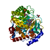
| ||||||||
|---|---|---|---|---|---|---|---|---|---|
| 1 |
| ||||||||
| Unit cell |
|
-Components
-Protein , 1 types, 1 molecules A
| #1: Protein | Mass: 39984.664 Da / Num. of mol.: 1 / Fragment: TRUNCATED Source method: isolated from a genetically manipulated source Source: (gene. exp.) Homo sapiens (human) / Gene: DHODH / Production host:  References: UniProt: Q02127, dihydroorotate dehydrogenase (quinone) |
|---|
-Non-polymers , 8 types, 277 molecules 


| #2: Chemical | ChemComp-FMN /  Mass: 456.344 Da / Num. of mol.: 1 / Source method: obtained synthetically / Formula: C17H21N4O9P Mass: 456.344 Da / Num. of mol.: 1 / Source method: obtained synthetically / Formula: C17H21N4O9P | ||||||||
|---|---|---|---|---|---|---|---|---|---|
| #3: Chemical | ChemComp-ORO /  Mass: 156.096 Da / Num. of mol.: 1 / Source method: obtained synthetically / Formula: C5H4N2O4 Mass: 156.096 Da / Num. of mol.: 1 / Source method: obtained synthetically / Formula: C5H4N2O4 | ||||||||
| #4: Chemical | ChemComp-VU7 /  Mass: 404.409 Da / Num. of mol.: 1 / Source method: obtained synthetically / Formula: C24H18F2N2O2 / Feature type: SUBJECT OF INVESTIGATION Mass: 404.409 Da / Num. of mol.: 1 / Source method: obtained synthetically / Formula: C24H18F2N2O2 / Feature type: SUBJECT OF INVESTIGATION | ||||||||
| #5: Chemical | ChemComp-ACT /  Mass: 59.044 Da / Num. of mol.: 4 / Source method: obtained synthetically / Formula: C2H3O2 Mass: 59.044 Da / Num. of mol.: 4 / Source method: obtained synthetically / Formula: C2H3O2#6: Chemical | ChemComp-SO4 /  Mass: 96.063 Da / Num. of mol.: 4 / Source method: obtained synthetically / Formula: SO4 Mass: 96.063 Da / Num. of mol.: 4 / Source method: obtained synthetically / Formula: SO4#7: Chemical |  Mass: 92.094 Da / Num. of mol.: 2 / Source method: obtained synthetically / Formula: C3H8O3 Mass: 92.094 Da / Num. of mol.: 2 / Source method: obtained synthetically / Formula: C3H8O3#8: Chemical | ChemComp-PGE / |  Mass: 150.173 Da / Num. of mol.: 1 / Source method: obtained synthetically / Formula: C6H14O4 Mass: 150.173 Da / Num. of mol.: 1 / Source method: obtained synthetically / Formula: C6H14O4#9: Water | ChemComp-HOH / | Mass: 18.015 Da / Num. of mol.: 263 / Source method: isolated from a natural source / Formula: H2O |
-Details
| Has ligand of interest | Y |
|---|
-Experimental details
-Experiment
| Experiment | Method: X-RAY DIFFRACTION / Number of used crystals: 1 |
|---|
- Sample preparation
| Crystal | Density Matthews: 3.53 Å3/Da / Density % sol: 65.2 % |
|---|---|
| Crystal grow | Temperature: 293 K / Method: vapor diffusion Details: 0.10 M NaAcetate pH 4.8, 2.30 M (NH4)2SO4, 30 % Glycerol, 18% (w/v) PEG 4000, 0.1M Na3Citrate pH5.75 |
-Data collection
| Diffraction | Mean temperature: 100 K / Serial crystal experiment: N |
|---|---|
| Diffraction source | Source: SYNCHROTRON / Site: SLS  / Beamline: X10SA / Wavelength: 1.00002 Å / Beamline: X10SA / Wavelength: 1.00002 Å |
| Detector | Type: DECTRIS PILATUS 6M / Detector: PIXEL / Date: Jan 9, 2019 |
| Radiation | Protocol: SINGLE WAVELENGTH / Monochromatic (M) / Laue (L): M / Scattering type: x-ray |
| Radiation wavelength | Wavelength: 1.00002 Å / Relative weight: 1 |
| Reflection | Resolution: 1.73→78.47 Å / Num. obs: 59048 / % possible obs: 96.2 % / Redundancy: 5.7 % / Biso Wilson estimate: 28.536 Å2 / Rmerge(I) obs: 0.064 / Rrim(I) all: 0.07 / Χ2: 0.997 / Net I/σ(I): 17.96 |
| Reflection shell | Resolution: 1.73→1.98 Å / Redundancy: 5.7 % / Rmerge(I) obs: 0.499 / Mean I/σ(I) obs: 4.01 / Num. unique obs: 19827 / Rrim(I) all: 0.549 / % possible all: 98.2 |
- Processing
| Software |
| |||||||||||||||||||||||||||||||||||||||||||||||||||||||||||||||||||||||||||
|---|---|---|---|---|---|---|---|---|---|---|---|---|---|---|---|---|---|---|---|---|---|---|---|---|---|---|---|---|---|---|---|---|---|---|---|---|---|---|---|---|---|---|---|---|---|---|---|---|---|---|---|---|---|---|---|---|---|---|---|---|---|---|---|---|---|---|---|---|---|---|---|---|---|---|---|---|
| Refinement | Method to determine structure: MOLECULAR REPLACEMENT Starting model: NONE Resolution: 1.73→78.47 Å / Cor.coef. Fo:Fc: 0.965 / Cor.coef. Fo:Fc free: 0.961 / SU B: 3.179 / SU ML: 0.053 / SU R Cruickshank DPI: 0.0805 / Cross valid method: THROUGHOUT / σ(F): 0 / ESU R: 0.08 / ESU R Free: 0.077 / Stereochemistry target values: MAXIMUM LIKELIHOOD
| |||||||||||||||||||||||||||||||||||||||||||||||||||||||||||||||||||||||||||
| Solvent computation | Ion probe radii: 0.8 Å / Shrinkage radii: 0.8 Å / VDW probe radii: 1.2 Å / Solvent model: MASK | |||||||||||||||||||||||||||||||||||||||||||||||||||||||||||||||||||||||||||
| Displacement parameters | Biso max: 93.64 Å2 / Biso mean: 26.958 Å2 / Biso min: 11.84 Å2
| |||||||||||||||||||||||||||||||||||||||||||||||||||||||||||||||||||||||||||
| Refinement step | Cycle: final / Resolution: 1.73→78.47 Å
| |||||||||||||||||||||||||||||||||||||||||||||||||||||||||||||||||||||||||||
| Refine LS restraints |
| |||||||||||||||||||||||||||||||||||||||||||||||||||||||||||||||||||||||||||
| LS refinement shell | Resolution: 1.73→1.775 Å
| |||||||||||||||||||||||||||||||||||||||||||||||||||||||||||||||||||||||||||
| Refinement TLS params. | Method: refined / Refine-ID: X-RAY DIFFRACTION
| |||||||||||||||||||||||||||||||||||||||||||||||||||||||||||||||||||||||||||
| Refinement TLS group |
|
